Early detection of ectopic pregnancy by ultrasound can save lives. Learn how ultrasound identifies ectopic pregnancies, symptoms to watch for, and treatment options.
By Shubhra Mishra — a mom of two who turned her own confusion during pregnancy into BumpBites, a global mission to make food choices clear, safe, and stress-free for every expecting mother. 💛
Check whether any food is safe during pregnancy with the BumpBites Food Safety Checker.
Quick take: Ultrasound is the frontline tool for spotting an ectopic pregnancy early. A transvaginal scan can usually see a misplaced gestational sac by the fifth to sixth week, and its accuracy climbs to over 90 % when performed by an experienced sonographer. If you have pain, spotting, or an abnormal rise in hCG, ask for an urgent ultrasound—early detection can mean a simple medical treatment instead of emergency surgery.
It’s 2 a.m., you’ve just felt a sharp cramp and a few drops of spotting after a positive pregnancy test. Your heart races as you wonder: “Is this a miscarriage? Or could it be something more dangerous?” You’re not alone—many expecting parents experience that exact moment of uncertainty. The good news is that modern ultrasound can answer the question quickly and safely, often before symptoms worsen.
In this guide we’ll walk through everything you need to know about ectopic pregnancy by ultrasound early detection. We’ll explain what an ectopic pregnancy is, why catching it early matters, how different types of scans work, what the images look like, and what to expect during the exam. We’ll also cover treatment options, risk‑reduction strategies, and the most common follow‑up questions you might have after the scan.
By the end of the article you’ll have a clear picture of the role ultrasound plays, the timeline of detection, and the next steps if your scan shows something unexpected. Let’s turn that worry into knowledge, so you can feel confident when you talk to your provider.
What is the role of ultrasound in detecting ectopic pregnancy?
An ectopic pregnancy occurs when a fertilized egg implants outside the uterine cavity—most often in the fallopian tube, but also in the ovary, cervix, or abdominal cavity. Because the tube cannot stretch like the uterus, the embryo quickly outgrows its space, leading to pain, internal bleeding, and potentially life‑threatening rupture.
Early detection is the cornerstone of safe management. Ultrasound provides a real‑time, non‑invasive view of where the pregnancy is located, allowing clinicians to differentiate a normal intrauterine gestation from an ectopic one. The scan can also assess the viability of the pregnancy, the amount of free fluid (a sign of possible rupture), and guide treatment decisions—whether medical (methotrexate) or surgical (laparoscopy).
Guidelines from the American College of Obstetricians and Gynecologists (ACOG) and the UK's National Institute for Health and Care Excellence (NICE) both recommend ultrasound as the first‑line imaging modality when an ectopic pregnancy is suspected, especially in the presence of pain, vaginal bleeding, or atypical hCG trends.
In practice, the ultrasound serves three main purposes:
Localization: confirming whether a gestational sac is inside the uterine cavity.
Assessment: evaluating the size of the ectopic mass and checking for free fluid.
Monitoring: following the response to medical therapy if methotrexate is chosen.
Because the scan is painless, widely available, and can be performed in a clinic setting, it remains the most reliable tool for early ectopic pregnancy detection. Moreover, the visual confirmation of an empty uterus or an abnormal adnexal mass helps clinicians avoid unnecessary surgery and tailor care to preserve future fertility whenever possible. This immediate insight is invaluable for both diagnosis and patient peace of mind, offering clarity during a stressful time.
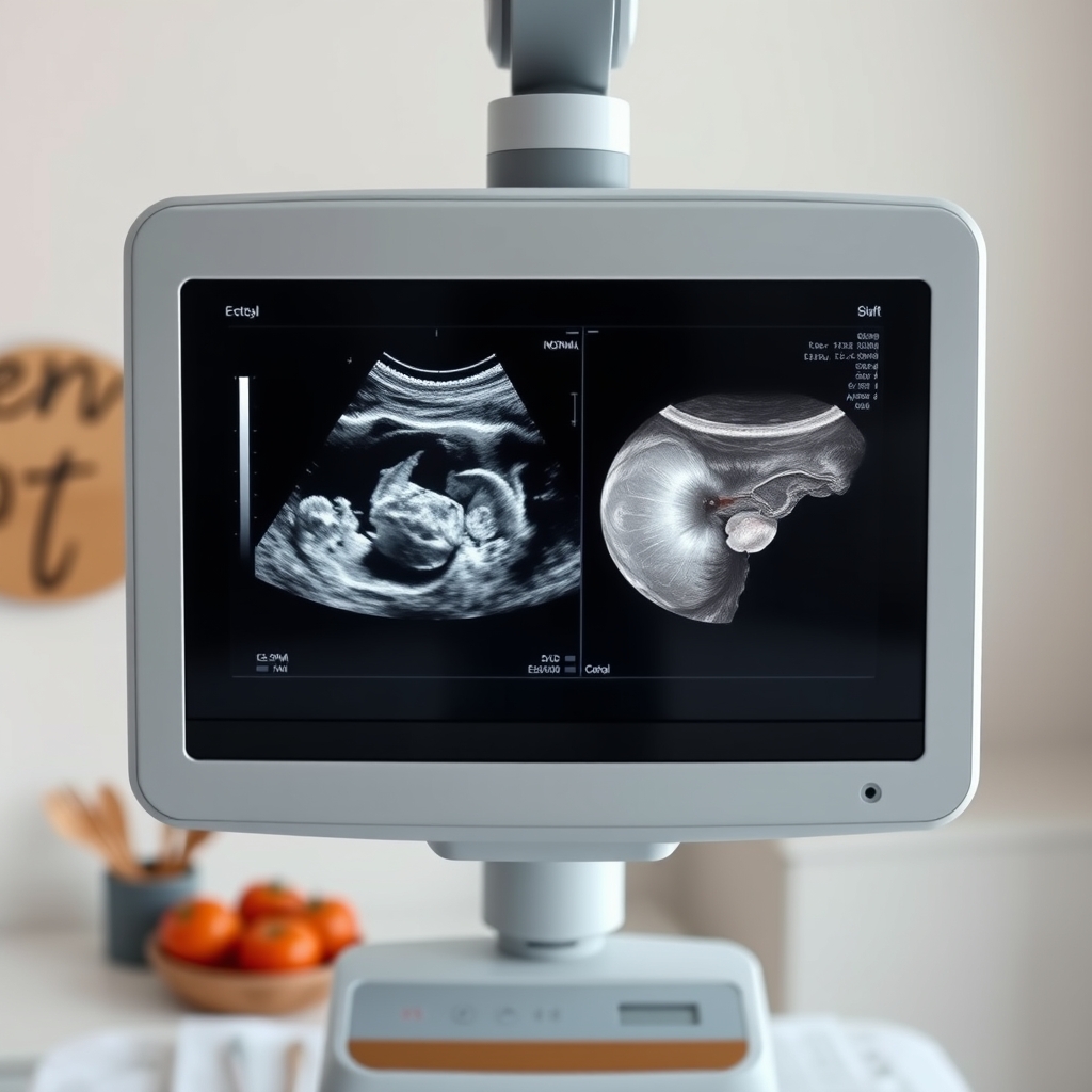
Typical transvaginal ultrasound view of an early ectopic pregnancy.
Early signs of ectopic pregnancy detected by ultrasound
When an ectopic pregnancy is still very early—around five weeks gestation—the ultrasound may not yet show a clearly defined sac. Instead, sonographers look for subtle clues that suggest an abnormal location.
Key early ultrasound findings include:
Empty uterus: No intrauterine gestational sac despite a positive pregnancy test and rising hCG.
Adnexal mass: A small, round or irregular structure near the ovary, sometimes described as a “tubal ring” sign.
Free fluid: Small pockets of fluid in the cul‑de‑sac (the pouch behind the uterus) can hint at a leaking tube.
Complex mass: A mixture of solid and cystic components that may contain a yolk sac without a fetal pole.
At five weeks, hCG levels typically range from 1,000 to 5,000 mIU/mL. If the level is above 1,500 mIU/mL and the uterus appears empty, the sensitivity of ultrasound for detecting an ectopic pregnancy jumps to about 70 % (ACOG 2023). By six weeks, the same hCG threshold yields a detection rate of roughly 90 %.
These early signs are why clinicians often repeat the scan 48–72 hours later if the first exam is inconclusive. The extra time can allow the sac to grow enough to become visible, or for fluid to accumulate enough to be seen. In practice, many patients find the repeat scan reassuring because it either confirms a normal intrauterine pregnancy or clarifies the need for further intervention. Sonographers also meticulously check for subtle findings, differentiating them from normal uterine changes or pseudo-sacs.
The “tubal ring” sign is a classic early indicator of ectopic implantation.
How accurate is ultrasound in diagnosing ectopic pregnancy?
Accuracy depends on several variables: gestational age, hCG level, sonographer expertise, and the type of ultrasound equipment used. Large, modern machines with high‑frequency probes improve resolution, especially for transvaginal scans.
According to a meta‑analysis of 22 studies published in the Journal of Ultrasound in Medicine, overall sensitivity for transvaginal ultrasound in detecting ectopic pregnancy is 93 % and specificity is 96 % when performed by experienced operators. Sensitivity drops to about 70 % for transabdominal scans in the first trimester because the probe is farther from the pelvic organs.
False‑negative results—where an ectopic pregnancy is missed—are most common when hCG levels are low (<1,000 mIU/mL) or when the ectopic is located high in the abdomen. False‑positives can arise from misinterpreting a corpus luteum cyst as an ectopic mass; however, distinguishing features such as peripheral blood flow patterns (seen on Doppler) help reduce this error.
Overall, when combined with clinical assessment (pain, bleeding, hCG trends), ultrasound reaches a diagnostic accuracy of over 95 % for most patients. This is why guidelines emphasize that a single “empty uterus” finding should not be the sole determinant; it must be interpreted in the full clinical context. While highly accurate, limitations arise with very small ectopics, unusual locations, or complex medical histories. Serial scans and hCG monitoring are often used in conjunction to ensure a definitive diagnosis and minimize false positives or negatives.
Can a transvaginal ultrasound detect ectopic pregnancy?
Yes—transvaginal ultrasound (TVUS) is the gold‑standard for early ectopic detection. The probe is inserted into the vagina, bringing the transducer within a few centimeters of the ovaries and fallopian tubes. This proximity yields images with a resolution as fine as 0.5 mm, far superior to the 5–7 mm resolution of a transabdominal scan.
Guidelines from the Royal College of Obstetricians and Gynaecologists (RCOG) recommend TVUS as the first‑line imaging technique when a patient presents with pelvic pain, vaginal bleeding, or an abnormal hCG rise in early pregnancy. The procedure is brief (usually under 10 minutes) and most women describe it as uncomfortable rather than painful.
Key advantages of TVUS include:
Earlier visualization of the gestational sac (as early as 4.5–5 weeks).
Better assessment of adnexal structures, allowing detection of the “tubal ring” sign.
Ability to use Doppler to evaluate blood flow, differentiating an ectopic from a corpus luteum cyst.
In rare cases where TVUS is contraindicated (e.g., severe vaginal bleeding), a high‑resolution transabdominal scan can still provide useful information, but the detection window shifts later to about 6–7 weeks. Patients typically lie on their back, and the probe is gently inserted. While it can feel slightly invasive, it's generally quick and well-tolerated. This proximity allows for clearer images, especially for subtle adnexal masses or small amounts of free fluid.
What are the symptoms of ectopic pregnancy that require ultrasound?
Symptoms alone cannot confirm an ectopic pregnancy, but they are the trigger that prompts an urgent scan. The most common warning signs include:
Pain: Sharp or crampy unilateral pelvic pain, often worsening with movement.
Vaginal bleeding: Light spotting or heavier bleeding that may be mistaken for a period.
Shoulder pain: Referred pain from intra‑abdominal blood irritating the diaphragm—a red flag for rupture.
Dizziness or faintness: Signs of internal bleeding or shock.
Abnormal hCG rise: hCG levels that rise slower than expected (less than 66 % increase over 48 hours) or plateau.
If you experience any of these, especially in combination, your provider will likely order an ultrasound right away. Even if pain is mild, an “empty uterus” on scan combined with these symptoms raises the suspicion high enough to warrant close monitoring. Pain can progress from a dull ache to sharp, stabbing, localized pain. Vaginal bleeding might be dark brown and intermittent, unlike a typical period. Shoulder pain, a critical symptom, arises from internal bleeding irritating the phrenic nerve.
How soon can an ectopic pregnancy be detected by ultrasound?
The detection window begins around 5 weeks of gestation, when the embryo measures roughly 2–3 mm. At this point, a transvaginal scan can often reveal the gestational sac if it is located in the tube. By 6 weeks, the sac typically reaches 5–6 mm, making it visible in almost all cases.
Below is a quick reference table that shows typical detection timing and corresponding hCG thresholds:
Gestational Age (weeks)
Typical hCG Level (mIU/mL)
Ultrasound Detection Rate (TVUS)
4.5–5
1,000–2,000
~70 %
5–5.5
2,000–3,500
~85 %
6–7
3,500–6,000
>90 %
7+
>6,000
~95 %
In practical terms, if you present with pain and a positive test at five weeks, a TVUS will most likely reveal an ectopic mass or confirm an empty uterus. If the first scan is inconclusive, a repeat scan in 48 hours is standard practice. This short‑interval follow‑up balances the need for timely diagnosis with the desire to avoid unnecessary radiation or invasive procedures. The hCG discriminatory zone (e.g., 1500-2000 mIU/mL for TVUS) is when an intrauterine sac *should* be visible. If hCG is above this with an empty uterus, an ectopic is suspected. The 48-72 hour wait allows for sufficient growth or clearer visualization.
Ultrasound images of ectopic pregnancy vs normal pregnancy
Seeing the difference on an ultrasound can be reassuring for both patients and clinicians. A normal intrauterine pregnancy at 6 weeks typically shows a gestational sac within the uterine cavity, a yolk sac, and possibly a tiny fetal pole with cardiac activity.
In contrast, an ectopic pregnancy image often displays:
Adnexal mass: A round or irregular echo‑dark structure near the ovary.
No intrauterine sac: The uterus appears empty despite a positive hCG.
Free fluid: Anechoic (dark) areas in the cul‑de‑sac indicating blood.
Doppler flow: A “ring of fire” pattern—intense peripheral blood flow around the mass.
Below is a side‑by‑side illustration (image placeholders) that highlights these differences. The left panel shows a healthy uterine sac; the right panel displays a classic tubal ectopic with surrounding fluid.
Normal pregnancy versus ectopic pregnancy on a transvaginal scan.
Understanding these visual cues helps patients feel more informed when their provider explains the findings. It also underscores why a skilled sonographer is essential—subtle differences can change management dramatically. The "ring of fire" on Doppler, showing increased blood flow around the mass, is a strong indicator. A normal sac is round/oval, centrally within the uterus, and grows predictably, often showing a fetal pole.
What to expect during an ultrasound for suspected ectopic pregnancy
First, your provider will review your symptoms and hCG results. You’ll be asked to lie on an exam table with a full bladder (for transabdominal views) or an empty bladder (for transvaginal scanning). The technician will apply a warm gel to the abdomen or gently insert a slim probe into the vagina.
The scan itself usually lasts 10–15 minutes. The sonographer will examine:
The uterus for an intrauterine sac.
The ovaries and fallopian tubes for any abnormal masses.
The cul‑de‑sac for free fluid.
Blood flow patterns using color Doppler.
During the exam, you may feel a mild pressure but no pain. It’s normal to hear the machine’s soft whirring and to see gray‑scale images on the monitor. After the scan, the provider will discuss the findings with you. If an ectopic pregnancy is confirmed, they’ll outline treatment options, which may include medication (methotrexate), minimally invasive surgery (laparoscopy), or, in rare emergencies, a laparotomy.
If the scan is inconclusive, expect a repeat scan in a couple of days, possibly combined with serial hCG measurements to track how quickly the hormone is rising or falling. Many clinics coordinate this follow‑up automatically, so you won’t have to chase the appointment yourself. The sonographer obtains images and measurements. While they cannot diagnose, they relay findings to your doctor, who will discuss them. It's important to ask questions and have support, as waiting for results can be emotionally challenging.
From our medical team: An early ultrasound can be both diagnostic and reassuring. Even when the image shows an “empty uterus,” the combination of hCG trends and clinical symptoms guides the next steps. Most ectopic pregnancies identified before rupture are successfully treated with a single dose of methotrexate, sparing you from surgery and preserving future fertility.
How is methotrexate monitored after an ectopic pregnancy?
If a methotrexate regimen is chosen, the medication works by stopping the rapidly dividing cells of the ectopic tissue. The FDA has approved methotrexate for this indication, and ACOG recommends weekly monitoring of hCG until the level drops below 5 mIU/mL. Typically, hCG is measured on days 1, 4, and 7 after the injection, then weekly thereafter.
A declining hCG curve—usually a 15 % drop between days 4 and 7—is a good sign that the ectopic tissue is resolving. If the decline is slower, clinicians may repeat the dose or consider surgical removal. The follow‑up ultrasound, usually performed 1–2 weeks after treatment, confirms that the adnexal mass has disappeared and that there is no residual fluid.
Patients should avoid alcohol, high‑dose folic acid, and NSAIDs while on methotrexate, as these can interfere with the drug’s effectiveness. The medical team will provide a written checklist of contraindicated substances and a timeline for safe return to normal activities. Patients may experience nausea, abdominal pain, and fatigue. Follow all post-treatment instructions, including avoiding strenuous activity and sexual intercourse until hCG is undetectable (2-4 weeks). Your team will guide recovery.
Typical hCG drop after a single dose of methotrexate.
Which risk factors increase the chance of an ectopic pregnancy?
Understanding personal risk helps you and your provider decide how closely to monitor early pregnancy. The most common factors include:
Previous ectopic pregnancy (raises risk to ~10 %).
Pelvic inflammatory disease or sexually transmitted infections that scar the tubes.
Tubal surgery or sterilization procedures that alter anatomy.
Smoking—nicotine impairs tubal motility and increases ectopic risk.
Assisted reproductive technologies (IVF, ICSI) that introduce embryos directly into the uterus but can still result in tubal implantation.
Age over 35 and a history of infertility treatments.
The NHS recommends that anyone with these risk factors receive an early hCG test and, if positive, an early transvaginal scan—often as soon as 5 weeks. Lifestyle modifications, such as quitting smoking and treating any sexually transmitted infections promptly, can lower the odds of a future ectopic pregnancy. Discussing your full medical history with a provider ensures that any red‑flags are caught before symptoms appear. PID causes scarring, blocking the egg's passage. Smoking impairs fallopian tube cilia, which move the egg, increasing risk of implantation outside the uterus. Discussing your full medical history is vital.
What happens if an ectopic pregnancy is left untreated?
If an ectopic pregnancy is not identified and treated promptly, it can lead to severe and life-threatening complications. As the embryo grows, it can cause the fallopian tube to rupture, leading to significant internal bleeding (hemorrhage). Symptoms of rupture include sudden, excruciating abdominal pain, shoulder pain, dizziness, and signs of shock. A ruptured ectopic pregnancy is a medical emergency requiring immediate surgery. Without swift treatment, it can be fatal, highlighting why early detection through ultrasound is critical for maternal safety. Seek urgent care if you suspect rupture.
Coping emotionally after an ectopic pregnancy
Discovering you have an ectopic pregnancy can be an incredibly difficult and emotionally complex experience. It often brings a mix of grief for the loss, fear for your health, and even trauma. It's normal to feel sadness, anger, and anxiety about future pregnancies. All these feelings are valid, and you don't have to navigate them alone. Seek support from your partner, trusted friends, family, or a support group for pregnancy loss. Consider speaking with a therapist specializing in reproductive grief. Allow yourself time to heal physically and emotionally, and don't hesitate to reach out for resources.
Future fertility and pregnancy after an ectopic
One of the most common questions after an ectopic pregnancy is about the possibility of future pregnancies. The good news is that most women who experience an ectopic pregnancy can go on to have successful intrauterine pregnancies. Your fertility after an ectopic depends on several factors, including the condition of your remaining fallopian tube(s) and whether a tube was removed. If one tube was preserved, your chances of conceiving again are generally good. However, the risk of a repeat ectopic pregnancy is higher, ranging from 10% to 15% (ACOG 2023). Because of this increased risk, it's crucial to work closely with your healthcare provider for early monitoring in any subsequent pregnancies. This typically involves early hCG testing and a transvaginal ultrasound as soon as a positive pregnancy test is confirmed to ensure the new pregnancy is safely located within the uterus.
Myth vs. fact
Myth: “If I have an ectopic pregnancy, I’ll always need surgery.”
Fact: About 70 % of early, unruptured ectopic pregnancies can be treated medically with methotrexate, provided hCG levels are below a certain threshold and the ectopic mass is smaller than 3.5 cm (ACOG 2022).
Myth: “A normal ultrasound means I’m definitely not pregnant.”
Fact: An empty uterus on a scan performed before 5 weeks does not rule out pregnancy. The gestational sac may simply be too small to see; repeat scanning or hCG monitoring is essential.
Myth: “Once I’ve had an ectopic pregnancy, I can’t get pregnant again.”
Fact: Most women can have a healthy subsequent pregnancy. The risk of another ectopic rises from ~1 % to about 10–15 % after one event, but careful early monitoring usually catches any repeat early.
Key takeaways
Ultrasound, especially transvaginal, is the most accurate early tool for spotting an ectopic pregnancy.
Symptoms like unilateral pelvic pain, spotting, or abnormal hCG trends should prompt an urgent scan.
Ectopic pregnancies are often visible by 5–6 weeks; a repeat scan may be needed if the first is inconclusive.
Medical treatment with methotrexate is possible for many early, unruptured ectopics, preserving fertility.
After an ectopic pregnancy, a normal early‑pregnancy ultrasound is recommended to confirm intrauterine implantation.
Call your provider immediately if you experience severe pain, heavy bleeding, shoulder pain, or faintness.
Frequently asked questions
What are the chances of having an ectopic pregnancy?
The overall risk is about 1–2 % of all pregnancies (CDC 2022). Certain factors—such as prior ectopic pregnancy, tubal surgery, or smoking—can raise that likelihood to 5–10 %.
Can an ectopic pregnancy be moved to the uterus?
Currently, there is no proven method to relocate an ectopic embryo safely. Treatment focuses on stopping the growth (methotrexate) or removing the tissue surgically.
How common is ectopic pregnancy?
In the United States, approximately 6–8 out of every 1,000 pregnancies are ectopic (ACOG 2023). It remains the leading cause of first‑trimester maternal death.
What causes an ectopic pregnancy?
Most cases result from tubal damage that impedes the embryo’s passage to the uterus. Causes include pelvic inflammatory disease, prior tubal surgery, endometriosis, and certain assisted‑reproductive techniques.
Can you have a normal pregnancy after an ectopic pregnancy?
Yes. Over 80 % of women who experience one ectopic pregnancy go on to have a healthy intrauterine pregnancy, especially with early monitoring and timely care.
What are the symptoms of a ruptured ectopic pregnancy?
Sudden, severe abdominal or shoulder pain, faintness, rapid heartbeat, and heavy vaginal bleeding signal a possible rupture. This is a medical emergency—call emergency services right away.
What should I do if my hCG levels are not rising as expected?
A slower‑than‑expected rise (less than a 66 % increase over 48 hours) is a red flag for ectopic or failing intrauterine pregnancy. Your provider will likely order an urgent ultrasound and repeat hCG tests to clarify the situation.
Can an ectopic pregnancy be seen on a home pregnancy test?
Home pregnancy tests detect hCG, not the location of the pregnancy. A positive test tells you you’re pregnant, but it cannot differentiate an intrauterine from an ectopic pregnancy. Ultrasound is required for that distinction.
What pain relief can I take for ectopic pregnancy discomfort?
For mild pain, your doctor may recommend acetaminophen (Tylenol). Avoid NSAIDs like ibuprofen, especially if you're receiving methotrexate, as they can interfere with the medication. Always consult your provider before taking any pain relievers during a suspected or diagnosed ectopic pregnancy to ensure it's safe and won't complicate your treatment plan.
Are there different types of ectopic pregnancies?
While most ectopic pregnancies occur in the fallopian tube (tubal pregnancy), they can implant in other locations. These include cervical (in the cervix), ovarian (on the ovary), abdominal (in the abdominal cavity), or interstitial (in the part of the fallopian tube that passes through the uterine wall). Each type requires specific diagnostic and management due to unique risks.
When to call your doctor
If you experience any of the following, seek immediate medical attention: sudden sharp pelvic pain, especially on one side; heavy or persistent vaginal bleeding; shoulder pain; dizziness, faintness, or rapid heartbeat; or a sudden drop in blood pressure. Remember, this article is for information only and does not replace personalized medical advice.
References
American College of Obstetricians and Gynecologists. “Management of Ectopic Pregnancy.” Practice Bulletin No. 193, 2023.
National Institute for Health and Care Excellence. “Ectopic Pregnancy: Diagnosis and Initial Management.” NICE Guideline NG111, 2022.
Royal College of Obstetricians and Gynaecologists. “Ectopic Pregnancy.” RCOG Green‑top Guideline No. 64, 2022.
Centers for Disease Control and Prevention. “Ectopic Pregnancy Surveillance.” CDC, 2022.
World Health Organization. “Recommendations for the Prevention and Management of Ectopic Pregnancy.” WHO, 2021.
Journal of Ultrasound in Medicine. “Meta‑analysis of Ultrasound Accuracy in Early Ectopic Pregnancy.” 2022.
British Pregnancy Advisory Service. “Understanding hCG Trends in Early Pregnancy.” BPA, 2023.
Mayo Clinic. “Ectopic Pregnancy.” Mayo Clinic, 2024.
U.S. Food and Drug Administration. “Methotrexate Injection: FDA‑approved uses and prescribing information.” FDA, 2023.
National Health Service (UK). “Early pregnancy: symptoms and when to seek help.” NHS, 2023.
Not sure about the label on Ectopic Pregnancy By Ultrasound Early Detection products?
Snap the ingredients list and SafeFilter checks every ingredient for your stage — only 3 free scans this month, then you're locked until reset. Unlimited from $7/mo or $50/yr.
When Shubhra Mishra was expecting her first child in 2016, she was overwhelmed by conflicting food advice — one site said yes, another said never. By the time her second baby arrived in 2019, she realized millions of mothers face the same confusion.
That sparked a five-year journey through clinical nutrition papers, cultural diets, and expert conversations — all leading to BumpBites: a calm, compassionate space where science meets everyday motherhood.
Her long-term vision is to build a global community ensuring safe, supported, and free deliveriesfor every mother — because no woman should face pregnancy alone or uninformed. 🌿
🌍 Stand with mothers, shape safer guidance
Join a small circle of experts who review BumpBites articles so expecting parents everywhere can decide with confidence.