Fetal growth restriction (FGR) can be detected early using the cerebroplacental ratio (CPR). Learn how CPR identifies blood flow redistribution and improves prenatal outcomes.
By Shubhra Mishra — a mom of two who turned her own confusion during pregnancy into BumpBites, a global mission to make food choices clear, safe, and stress-free for every expecting mother. 💛
Check whether any food is safe during pregnancy with the BumpBites Food Safety Checker.
Download the Complete Pregnancy Food Guide (10,000 Foods) 📘
Instant PDF download • No spam • Trusted by thousands of moms
💡 Your email is 100% safe — no spam ever.
Quick take: A low cerebroplacental ratio (CPR) on Doppler ultrasound signals that blood flow is being redirected toward the brain—a hallmark of fetal growth restriction (FGR). It can appear before the umbilical artery shows problems, helping clinicians intervene earlier. Normal CPR values vary by gestational age, and values below the trimester‑specific cut‑off warrant closer monitoring and sometimes delivery planning.
It’s 2 a.m. and you’ve just finished a restless night of heart‑racing contractions, nausea, and a sudden, vague worry: “Did that ultrasound yesterday show anything abnormal?” You glance at the report, see the words “cerebroplacental ratio,” and wonder whether you’re looking at a red flag or a harmless number. You’re not alone—many expecting parents encounter CPR for the first time when their provider is checking for early signs of fetal growth restriction. In this guide we’ll demystify the cerebroplacental ratio, explain exactly how it’s measured, show you what “normal” looks like at each week of pregnancy, and outline what a low reading means for you and your baby.
By the end of this article you’ll know the physiological basis of CPR, the ultrasound technique behind it, the gestational‑age specific cut‑offs, and how CPR compares with other Doppler indices such as the umbilical artery (UA) and middle cerebral artery pulsatility index (MCA‑PI). We’ll also walk through management steps for a low CPR, discuss its predictive value for adverse outcomes, and flag the current limitations of the test. All of this is grounded in the latest guidance from ACOG, NICE, and the International Society of Ultrasound in Obstetrics and Gynecology (ISUOG).
What is the cerebroplacental ratio and why does it matter?
The cerebroplacental ratio (CPR) is a simple calculation that compares blood‑flow resistance in two fetal vessels: the middle cerebral artery (MCA) and the umbilical artery (UA). In healthy pregnancies, the UA—carrying deoxygenated blood from the fetus to the placenta—has a higher resistance than the MCA, which supplies oxygen‑rich blood to the brain. The CPR is expressed as:
CPR = MCA pulsatility index ÷ Umbilical artery pulsatility index
A higher CPR indicates that the brain is receiving a proportionally larger share of blood flow, which is normal. When the placenta begins to falter—often the first step in fetal growth restriction—the fetus initiates a protective mechanism called “brain‑sparring.” The brain vessels dilate, reducing the MCA resistance, while the placenta’s resistance may rise. The result is a lower CPR, signaling that the fetus is redistributing blood to protect its brain at the expense of other organs.
Because this redistribution can happen before any change in fetal size or the classic “absent end‑diastolic flow” in the UA, CPR serves as an early marker of FGR. Detecting it early gives clinicians a chance to monitor more closely, adjust maternal nutrition or medication, and plan delivery at the optimal time to minimize the risk of stillbirth or neonatal complications.
Beyond its role in identifying growth restriction, a low CPR has been linked in several cohort studies to neurodevelopmental outcomes later in childhood. While the evidence is still evolving, early detection allows for targeted interventions—such as optimizing maternal blood pressure or timing delivery—to mitigate potential long‑term effects.
Finally, CPR offers a window into fetal physiology that is not captured by size measurements alone, making it a valuable addition to routine third‑trimester surveillance when risk factors are present.
How is CPR measured on ultrasound?
Measuring CPR requires a standardized Doppler ultrasound exam performed by a qualified sonographer or obstetrician. The steps are:
Position the fetus. The examiner first visualizes the fetal head in a transverse (cross‑sectional) view, ideally with the brain midline perpendicular to the transducer.
Obtain the MCA waveform. Using a small sample volume (< 2 mm), the sonographer places the Doppler gate on the proximal MCA, just distal to its origin from the circle of Willis. The angle of insonation should be ≤ 30°, and the pulse‑repetition frequency (PRF) is set low enough to capture the low‑velocity flow.
Record the MCA pulsatility index (PI). The PI is calculated as (peak systolic velocity – end‑diastolic velocity) ÷ mean velocity. Most machines provide an automatic PI calculation; the operator should verify that the waveform has a clear triphasic pattern and that the end‑diastolic flow is not zero.
Switch to the umbilical artery. The probe is moved to the free loop of the cord, and the Doppler gate is placed on the UA close to the fetal insertion point. Again, the angle is kept ≤ 30°, and the PI is recorded.
Calculate the CPR. The ratio is derived by dividing the MCA‑PI by the UA‑PI. Many modern ultrasound systems can compute this automatically, but the manual calculation ensures consistency across machines.
It’s important that the fetus is not actively moving or that the mother is not breathing heavily, as motion can distort the waveforms. The entire CPR assessment typically takes 2–3 minutes, and no radiation or contrast agents are used—so the test is considered safe for both mother and baby.
Quality control is essential. The American College of Obstetricians and Gynecologists (ACOG) recommends that each Doppler study be reviewed for angle accuracy and for the presence of a clean, low‑noise signal before the ratio is recorded. When a result is borderline, a repeat scan within 24–48 hours helps confirm that the finding reflects true physiology rather than transient fetal movement or technical variance.
Because the procedure is brief and non‑invasive, most women can have CPR measured during a routine anatomy scan without needing additional appointments, which helps integrate it smoothly into standard prenatal care.
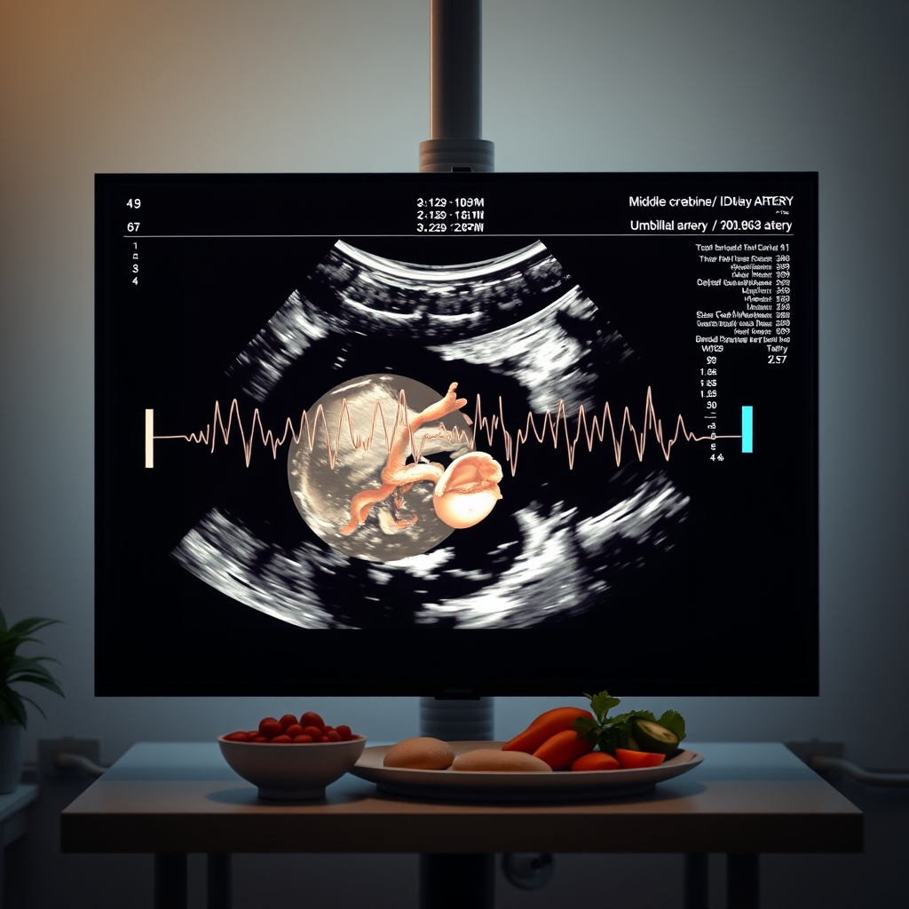
During a routine scan, the sonographer captures Doppler waveforms from the MCA and UA to calculate the CPR.
Normal CPR ranges and gestational‑age specific cut‑offs
Because both MCA‑PI and UA‑PI naturally change as the pregnancy progresses, CPR norms are expressed as gestational‑age‑specific percentiles. Large reference studies, such as those compiled by the International Society of Ultrasound in Obstetrics and Gynecology (ISUOG) and the Society for Maternal‑Fetal Medicine (SMFM), provide the following approximate median CPR values and 5th‑percentile cut‑offs:
Gestational Age (weeks)
Median CPR
5th‑percentile (low‑risk cut‑off)
20‑22
1.30
0.95
24‑26
1.25
0.90
28‑30
1.20
0.85
32‑34
1.15
0.80
36‑38
1.10
0.75
These numbers are not absolute thresholds; they are meant as guidance. In practice, many clinicians use a CPR < 0.8 as a “red flag” regardless of gestational age, especially when paired with other concerning findings such as abnormal umbilical artery Doppler or a fetal abdominal circumference below the 10th percentile.
National health services, including the NHS in the United Kingdom, have incorporated locally derived reference charts that adjust for maternal body‑mass index (BMI) and fetal sex, refining the cut‑offs for diverse populations. When you’re reviewing your own report, look for a footnote indicating which reference set was used.
If you’d like to calculate your own CPR based on the most recent ultrasound measurements, you can use the Cerebroplacental Ratio (CPR) calculator on our site. It will automatically apply the appropriate gestational‑age reference ranges and tell you whether your value falls within the normal range.
Remember that reference ranges can shift slightly between institutions, so a “low” CPR in one clinic may still be within the normal range in another that uses a different population‑based chart.
CPR versus other Doppler indices
Historically, the umbilical artery PI has been the cornerstone of Doppler surveillance for FGR. More recently, the MCA‑PI and combined CPR have gained prominence because they capture the fetal compensatory response, not just placental resistance. Below is a quick comparison:
Umbilical artery PI (UA‑PI): Detects increased placental resistance. Absent or reversed end‑diastolic flow is a late sign of severe placental insufficiency.
Middle cerebral artery PI (MCA‑PI): Reflects cerebral vasodilation. A low MCA‑PI alone can indicate brain‑sparring but may be missed if the UA is still normal.
CPR: Integrates both measurements, offering a single ratio that flags early redistribution even when UA‑PI is still within normal limits.
Recent meta‑analyses cited by ACOG and NICE show that adding CPR to a standard UA‑PI protocol improves prediction of adverse perinatal outcomes by roughly 10‑15 %. Moreover, combined indices that include uterine artery Doppler (a measure of maternal placental perfusion) with CPR are emerging as even stronger predictors, especially for early‑onset FGR.
In practice, a low CPR often prompts clinicians to add a uterine artery Doppler study and a more frequent biophysical profile schedule, creating a comprehensive picture of both fetal and maternal circulatory health.
Because each index reflects a different physiological compartment, using them together reduces false‑positive alarms and helps tailor surveillance intensity to the individual pregnancy.
What a low CPR tells us – early signs of fetal growth restriction
A low CPR is essentially an early alarm bell that the fetus is redirecting blood toward the brain. This redistribution can precede measurable reductions in fetal size, which means that CPR can be the first objective clue that a baby is not growing as expected. Common scenarios in which a low CPR appears include:
Maternal hypertension or pre‑eclampsia, where placental perfusion is compromised.
Smoking or high‑altitude residence, both of which can affect oxygen delivery.
Maternal anemia or malnutrition, leading to reduced oxygen‑carrying capacity.
Previous history of FGR, prompting early and serial Doppler surveillance.
When a low CPR is identified, clinicians typically increase the frequency of surveillance. This may involve weekly biophysical profiles, more detailed growth scans, and sometimes fetal heart‑rate monitoring (NST) to watch for decelerations that would indicate worsening hypoxia.
Evidence from the SMFM consensus statement indicates that a CPR below the 5th percentile is associated with a two‑fold increase in stillbirth risk compared with fetuses whose CPR remains above that threshold, even after adjusting for maternal age and BMI. This statistic underscores why early detection matters: timely intervention can shift outcomes from emergency delivery to planned, controlled birth.
In addition, a low CPR can help clinicians differentiate between “true” growth restriction and constitutionally small but healthy fetuses, guiding more nuanced counseling for expectant parents.
Seeing a normal CPR can bring peace of mind, even when other signs are borderline.
Managing a low CPR in FGR – guidelines and next steps
When a low CPR is detected, the management plan depends on gestational age, the degree of reduction, and any accompanying Doppler abnormalities. Below is a practical algorithm that aligns with ACOG Practice Bulletin 2023 and NICE NG153 recommendations:
Confirm the result. Repeat the CPR in 24–48 hours to rule out technical error or transient fetal movement effects.
Assess growth. Measure abdominal circumference (AC) and estimate fetal weight. If the AC is < 10th percentile, the case is classified as FGR.
Stratify risk.
Early‑onset (≤ 32 weeks) with CPR < 0.7: Admit for close monitoring, consider corticosteroids for lung maturity, and plan delivery if UA‑PI becomes abnormal or if fetal distress emerges.
Late‑onset (> 32 weeks) with CPR < 0.8: Increase surveillance to twice weekly, perform non‑stress tests, and discuss timing of delivery if there are signs of worsening placental function.
Address modifiable factors. Counsel on smoking cessation, optimize maternal nutrition, and treat hypertension according to ACOG guidelines.
Consider adjunct therapies. Some centers use low‑dose aspirin (81 mg) for high‑risk pregnancies, though evidence for benefit after a low CPR has been identified is still emerging.
Plan delivery. If the fetus reaches ≥ 34 weeks and the CPR remains low with stable UA‑PI, many providers will aim for delivery by 37 weeks to reduce stillbirth risk. Earlier delivery may be warranted if there’s rapid deterioration.
In addition to the steps above, many obstetric units now incorporate a “maternal‑fetal medicine round” where a multidisciplinary team reviews each low‑CPR case. This collaborative approach ensures that decisions about corticosteroid timing, magnesium sulfate administration (for neuroprotection in pre‑eclampsia), and neonatal team notification are coordinated.
Throughout this process, clear communication with the family is key. Explain that a low CPR does not guarantee a bad outcome, but it does mean the care team will watch more closely and may intervene sooner to protect the baby’s health.
When the pregnancy reaches term, the decision to deliver often balances the CPR trend against other maternal and fetal signals, aiming for the safest possible timing.
Limitations of CPR and areas for future research
While CPR is a valuable tool, it is not without drawbacks:
Operator dependency. Accurate Doppler angle and proper placement of the sample volume are critical; inexperienced sonographers may generate erroneous ratios.
Variability between machines. Different ultrasound manufacturers use slightly different algorithms for PI calculation, which can affect the absolute CPR number.
Population‑specific reference ranges. Most published norms are derived from Caucasian cohorts; data for diverse ethnic groups are still limited, potentially affecting the applicability of cut‑offs.
Overlap with normal variation. Some healthy fetuses naturally have CPR values near the 5th percentile without any adverse outcomes, leading to possible over‑monitoring.
Research is ongoing to address these gaps. Large, multi‑ethnic cohort studies are being designed to refine gestational‑age‑specific percentiles. Machine‑learning algorithms that integrate CPR with other biometric data (e.g., uterine artery Doppler, maternal serum markers) aim to improve predictive accuracy. Until such data become mainstream, clinicians rely on a combination of CPR, UA‑PI, fetal growth charts, and clinical context to make informed decisions.
Another emerging line of inquiry explores whether serial CPR measurements—tracking the trend rather than a single value—better predicts outcomes than a one‑time cut‑off. Early results suggest that a rapidly falling CPR trajectory may identify the highest‑risk fetuses, but larger trials are needed before guidelines can be updated.
Finally, ongoing work is evaluating how CPR might be combined with novel biomarkers such as placental growth factor (PlGF) to create a composite risk score that could guide therapy more precisely.
Underlying causes of fetal growth restriction
Fetal growth restriction (FGR) is a multifactorial condition, and CPR is only a window into one piece of the puzzle. The most common driver is placental insufficiency, where the placenta cannot deliver enough oxygen and nutrients to match the fetus’s metabolic demands. This can stem from abnormal spiral‑artery remodeling early in pregnancy, leading to reduced uteroplacental blood flow.
Maternal health conditions also play a big role. Chronic hypertension, pre‑eclampsia, diabetes (especially when poorly controlled), and autoimmune diseases such as antiphospholipid syndrome can all compromise placental function. Infections like cytomegalovirus or TORCH agents may directly affect fetal growth, while lifestyle factors—smoking, excessive alcohol, and severe malnutrition—further aggravate the risk.
Understanding the root cause helps clinicians decide whether an intervention (e.g., antihypertensive therapy, infection treatment, or nutritional supplementation) might improve placental performance and, consequently, the CPR trajectory.
When the underlying etiology is identified early, targeted therapies—like low‑dose aspirin for pre‑eclampsia risk or iron supplementation for anemia—can sometimes reverse or stabilize the abnormal CPR pattern.
Maternal lifestyle and CPR: nutrition, smoking, altitude
Several modifiable factors influence CPR values and overall fetal wellbeing. Smoking introduces carbon monoxide, which reduces fetal oxygen saturation and often leads to a lower CPR as the fetus attempts to protect its brain. Quitting smoking—even a few weeks before the ultrasound—has been shown to improve both UA‑PI and CPR measurements.
Maternal nutrition matters, too. Diets rich in iron, folate, and omega‑3 fatty acids support healthy placental development. Conversely, severe caloric restriction or micronutrient deficiencies can precipitate early brain‑sparring, reflected as a low CPR. Some clinicians recommend a balanced Mediterranean‑style diet for pregnant women at risk of FGR, citing NICE guidance on maternal nutrition.
Living at high altitude (> 2,500 m) naturally lowers ambient oxygen pressure, which can cause a physiologic reduction in fetal oxygen delivery. Studies from Andean and Himalayan populations demonstrate a shift toward lower CPR values at baseline, but also a higher threshold for what constitutes “abnormal.” If you reside at altitude, your provider may use altitude‑adjusted reference ranges to interpret the ratio accurately.
Hydration and regular prenatal exercise (as approved by your provider) also promote optimal blood flow, potentially stabilizing CPR values throughout the third trimester.
Emerging tools: AI and integrated biomarkers
Artificial intelligence is beginning to reshape how CPR data are interpreted. Researchers are training deep‑learning models on thousands of Doppler waveforms, combining CPR with uterine artery indices, maternal serum markers (like PAPP‑A), and clinical variables to produce individualized risk scores. Early pilot studies suggest that AI‑enhanced scores outperform CPR alone in predicting preterm delivery and neonatal intensive‑care admission.
While these technologies are not yet part of routine care, they illustrate a trend toward multimodal surveillance. In the near future, you may see an “integrated fetal health dashboard” that automatically pulls your latest CPR, growth measurements, and lab results into a single visual risk graphic—making it easier for both clinicians and families to understand the big picture.
Such dashboards could also flag when a single low CPR reading is likely a false alarm, reducing unnecessary anxiety and interventions.
Future dashboards may combine CPR with AI‑driven risk scores for clearer decision‑making.
From our medical team: A low CPR should be taken seriously—but not with panic. It tells us the fetus is adapting to a subtle placental problem, and that adaptation can be monitored and, in many cases, corrected with timely care. If your provider notes a low CPR, expect more frequent scans and possibly earlier delivery planning, but also expect reassurance that many babies with low CPR go on to have healthy outcomes when managed appropriately.
CPR in twin pregnancies
Twins present a unique hemodynamic environment because each fetus may share a placenta (monochorionic) or have separate placentas (dichorionic). CPR can be measured for each twin individually, but reference ranges are slightly lower than for singletons, reflecting the overall smaller size and different vascular demands of twins.
In monochorionic twins, twin‑to‑twin transfusion syndrome (TTTS) can dramatically alter Doppler patterns, making CPR an early warning sign of unequal blood sharing. Clinicians often combine CPR with middle cerebral artery peak systolic velocity (MCA‑PSV) to monitor for anemia in the donor twin and polycythemia in the recipient twin.
Because twin pregnancies carry a higher baseline risk of FGR, many obstetric units schedule Doppler surveillance—including CPR—every two weeks after 28 weeks, especially if growth discordance exceeds 20 %.
Using CPR to guide timing of delivery in pre‑eclampsia
Pre‑eclampsia creates a hostile placental environment, and CPR can help fine‑tune delivery timing when maternal blood pressure is controlled but fetal well‑being remains uncertain. A persistently low CPR (especially < 0.7) alongside a stable umbilical artery flow often prompts clinicians to consider delivery at 34–36 weeks, balancing the risks of prematurity against the danger of continued placental insufficiency.
Guidelines from ACOG and NICE advise that when CPR deteriorates rapidly, even in the absence of other severe Doppler changes, the threshold for delivery may be lowered to protect the brain and prevent stillbirth. This approach underscores how CPR adds nuance to the classic “blood pressure‑plus‑gestational age” decision matrix.
Myth vs. fact
Myth: A low CPR means the baby will definitely be small or have complications.
Fact: CPR is a risk marker, not a diagnosis. Many fetuses with a low ratio continue to grow normally, especially when the underlying cause (e.g., maternal hypertension) is controlled.
Myth: CPR testing harms the fetus because it uses “strong” ultrasound waves.
Fact: Doppler ultrasound uses low‑intensity sound waves that are considered safe for both mother and baby. The exam lasts only a few minutes and does not involve radiation.
Myth: Only specialists can interpret CPR, so it’s not useful in community clinics.
Fact: While expertise improves accuracy, most obstetric ultrasound machines now provide automated CPR calculations, allowing trained midwives and family physicians to incorporate the measure into routine prenatal care.
Key takeaways
CPR compares blood‑flow resistance in the fetal brain (MCA) and placenta (UA); a lower ratio signals early brain‑sparring.
Normal CPR values decline gradually across gestation; a value below the 5th percentile or < 0.8 generally warrants closer monitoring.
Measuring CPR is safe, quick, and performed during a standard Doppler ultrasound.
Low CPR can appear before any change in fetal size, making it a valuable early marker of fetal growth restriction.
Management involves repeat scans, growth assessment, maternal health optimization, and possibly earlier delivery if the situation deteriorates.
CPR should be interpreted alongside other Doppler indices and clinical factors; it is not a standalone diagnostic tool.
Emerging AI tools and integrated biomarkers may soon enhance CPR’s predictive power.
Frequently asked questions
What does a low CPR indicate in pregnancy?
A low CPR suggests that the fetus is redirecting blood toward the brain, a sign of early placental insufficiency and potential fetal growth restriction.
How is the cerebroplacental ratio calculated?
CPR is calculated by dividing the middle cerebral artery pulsatility index (MCA‑PI) by the umbilical artery pulsatility index (UA‑PI) obtained from a Doppler ultrasound.
Can CPR detect fetal growth restriction before other tests?
Yes. Because CPR reflects the fetus’s compensatory response, it can become abnormal weeks before a reduction in abdominal circumference or abnormal umbilical artery flow is seen.
What are the normal CPR values at 30 weeks gestation?
At 30 weeks, the median CPR is about 1.20, and the 5th‑percentile cut‑off (often used as a low‑risk threshold) is roughly 0.85.
What management steps are recommended for a low CPR?
Guidelines advise confirming the finding, assessing fetal growth, increasing surveillance (often twice weekly), addressing maternal risk factors, and considering early delivery if the ratio stays low or other Doppler abnormalities develop.
Is CPR measurement safe for the fetus?
Yes. Doppler ultrasound uses low‑energy sound waves, poses no known risk to the fetus, and the scan typically lasts only a few minutes.
Can I improve my CPR by changing my diet or lifestyle?
While diet alone won’t directly change the ratio, optimizing nutrition, staying hydrated, quitting smoking, and managing blood pressure can improve overall placental health, which may lead to a more favorable CPR on subsequent scans.
Is CPR useful in twin pregnancies?
CPR can be applied to each twin individually, but reference ranges differ slightly because twins often have lower birth weights. Clinicians usually interpret CPR alongside twin‑specific growth charts and consider the added complexity of shared placental circulation.
Can CPR be used after 40 weeks?
Yes. Even at term, a low CPR may signal that the placenta is no longer supporting the fetus adequately, prompting consideration of delivery if other signs of compromise are present.
Is there a difference between CPR and the cerebro‑umbilical ratio?
Both terms describe the same calculation—MCA‑PI divided by UA‑PI. “Cerebro‑umbilical ratio” is an older phrase; “cerebroplacental ratio” is now preferred in most guidelines.
When to call your doctor
If you notice any of the following, contact your obstetric provider immediately: sudden decrease in fetal movements, vaginal bleeding, severe abdominal pain, signs of pre‑eclampsia (headache, visual changes, swelling), or a new ultrasound report that shows a CPR below the recommended cut‑off. This article is for informational purposes only and does not replace personalized medical advice.
References
American College of Obstetricians and Gynecologists (ACOG). Practice Bulletin No. 226: Fetal Growth Restriction. 2023.
National Institute for Health and Care Excellence (NICE). NG153: Antenatal care for uncomplicated pregnancies. 2022.
International Society of Ultrasound in Obstetrics and Gynecology (ISUOG). Recommendations for Doppler assessment of fetal circulation. 2021.
Society for Maternal‑Fetal Medicine (SMFM). Consensus statement on the use of cerebroplacental ratio in fetal surveillance. 2022.
World Health Organization (WHO). Guidelines on antenatal care for a positive pregnancy experience. 2022.
Royal College of Obstetricians and Gynaecologists (RCOG). Fetal growth restriction: detection and management. 2021.
National Health Service (NHS). Doppler ultrasound in pregnancy: what to expect. 2023.
American Academy of Pediatrics (AAP). Neonatal outcomes after early‑onset fetal growth restriction. 2022.
When Shubhra Mishra was expecting her first child in 2016, she was overwhelmed by conflicting food advice — one site said yes, another said never. By the time her second baby arrived in 2019, she realized millions of mothers face the same confusion.
That sparked a five-year journey through clinical nutrition papers, cultural diets, and expert conversations — all leading to BumpBites: a calm, compassionate space where science meets everyday motherhood.
Her long-term vision is to build a global community ensuring safe, supported, and free deliveriesfor every mother — because no woman should face pregnancy alone or uninformed. 🌿
🌍 Stand with mothers, shape safer guidance
Join a small circle of experts who review BumpBites articles so expecting parents everywhere can decide with confidence.