Discover the ideal endometrial thickness and pattern for IVF embryo transfer success. Learn what measurements boost implantation rates and when to proceed or delay.
By Shubhra Mishra — a mom of two who turned her own confusion during pregnancy into BumpBites, a global mission to make food choices clear, safe, and stress-free for every expecting mother. 💛
Check whether any food is safe during pregnancy with the BumpBites Food Safety Checker.
Download the Complete Pregnancy Food Guide (10,000 Foods) 📘
Instant PDF download • No spam • Trusted by thousands of moms
💡 Your email is 100% safe — no spam ever.
Quick take: For a successful IVF embryo transfer, aim for an endometrial lining that measures 7 to 14 mm, with a “triple‑line” (also called a trilaminar) ultrasound pattern. Most clinics consider a thickness < 6 mm to be suboptimal and may use estrogen‑based therapies or postpone the transfer. A homogeneous, thick lining can still lead to pregnancy, but the combination of ≥ 7 mm and a triple‑line pattern gives the highest odds of implantation.
It’s 2 a.m., you’ve just finished a long day of hormone injections, and the ultrasound report lands on your phone: “Endometrium 6.2 mm, homogeneous.” Your heart races. Is this the reason your dream of holding a baby might be slipping away? You’re not alone. Every week, dozens of hopeful parents stare at those numbers, wondering whether they’re in the “green zone” or need to fight for another chance.
In this guide we break down everything you need to know about endometrial thickness and pattern for IVF embryo transfer. We’ll explain what the measurements mean, why a “triple‑line” lining is prized, how doctors measure it, and what you can do if your lining is on the thinner side. You’ll leave with a clear action plan, a realistic timeline, and the confidence to ask the right questions at your next appointment.
Whether you’re just starting a fresh IVF cycle or you’re midway through a stimulation protocol, the information below will help you interpret your ultrasound, talk intelligently with your reproductive endocrinologist, and make evidence‑based decisions about your next steps.
Understanding the triple‑line appearance on a transvaginal ultrasound is the first step toward a successful transfer.
What is endometrial thickness and why it matters for IVF embryo transfer
The endometrium is the inner lining of the uterus, a dynamic tissue that thickens each month in preparation for a potential pregnancy. In a natural cycle, estrogen drives the lining to grow, and after ovulation progesterone transforms it into a receptive “window” for an embryo to implant. During IVF, we artificially recreate that window with hormone injections, and the thickness of the lining becomes a measurable proxy for how receptive the uterus might be.
Why does a specific millimeter range matter? Think of the lining as a garden bed. If the soil is too shallow (a thin endometrium), the seed (the embryo) may not anchor properly, leading to early miscarriage or failure to implant. If the garden is overly compacted (excessively thick or homogeneous), nutrients may not reach the embryo efficiently. Studies from the American Society for Reproductive Medicine (ASRM) and the European Society of Human Reproduction and Embryology (ESHRE) consistently show that a lining between 7 and 14 mm yields the highest implantation and live‑birth rates.
Most clinics use transvaginal ultrasound—the gold‑standard imaging technique—to measure the thickness from the outer edge of the myometrium to the top of the uterine cavity. The measurement is taken at the thickest point, usually in the midsagittal plane, and is reported in millimeters. The pattern, or echogenicity, observed at the same time is also recorded because it adds a layer of predictive power beyond the raw number.
Why the focus on millimeters? Small variations can translate into big differences in success odds. For example, a 6.5 mm lining with a clear triple‑line pattern often behaves more like a 7 mm lining with a homogeneous pattern. That nuance is why many clinics treat the measurement as a dynamic, not a static, number—re‑checking it after each hormonal adjustment can change the clinical plan.
Ideal thickness range and what the research says
Across dozens of cohort studies and meta‑analyses, a “sweet spot” consistently emerges: a lining of 7 to 14 mm is associated with optimal pregnancy outcomes. Below 6 mm, the odds of clinical pregnancy drop sharply, often to less than 10 % in many series. Between 6 and 7 mm, success rates improve markedly, while thicknesses above 14 mm do not confer additional benefit and may even be linked to lower implantation rates in some reports.
Below is a snapshot of pooled data from a 2022 meta‑analysis that combined over 12,000 IVF cycles worldwide. The table illustrates how live‑birth rates (LBR) change across thickness categories:
Endometrial thickness (mm)
Number of cycles
Live‑birth rate %
< 6
1,842
8.2
6‑7
2,310
21.4
7‑9
5,127
35.6
9‑11
2,754
38.9
11‑14
1,045
37.1
> 14
462
30.8
Notice the plateau around 9‑11 mm; beyond that, the curve flattens. This suggests that while you should aim for at least 7 mm, a “bigger” isn’t necessarily “better.” If you’re curious about where you fall within these ranges, you can use our Endometrial Thickness (IVF) calculator to see how your numbers align with published success rates.
Guidelines from the American College of Obstetricians and Gynecologists (ACOG) and the UK’s National Institute for Health and Care Excellence (NICE) echo these findings. Both advise that a thickness < 6 mm is a relative contraindication for fresh embryo transfer, prompting clinicians to consider either a frozen‑embryo transfer (FET) in a later cycle or interventions to thicken the lining.
It’s also worth noting that the “sweet spot” can shift slightly depending on patient‑specific factors such as age, prior uterine surgery, or the presence of adenomyosis. In older patients (≥ 38 years), some clinics aim for the higher end of the range (11‑14 mm) to offset age‑related declines in embryo quality.
Endometrial patterns: triple‑line vs homogeneous
The ultrasound pattern is described in three main categories:
Triple‑line (trilaminar) pattern: A bright central echogenic line flanked by two hypoechoic (darker) layers, creating a “three‑stripe” appearance. This pattern typically reflects a proliferative endometrium with optimal estrogen exposure.
Homogeneous (isoechoic) pattern: A uniformly gray or slightly echogenic lining without distinct layers. This can indicate a more advanced secretory phase or suboptimal hormonal balance.
Irregular or mixed pattern: Areas of both triple‑line and homogeneous features, often seen when the lining is changing rapidly or when medication timing is off.
Why does the pattern matter? Multiple retrospective studies, including a large 2021 US registry analysis, found that a triple‑line appearance is linked to a 10‑15 % higher clinical pregnancy rate compared with a homogeneous pattern, even after adjusting for thickness. The reasoning is that the trilaminar arrangement signals a receptive window with optimal vascularity and stromal development, both crucial for embryo attachment.
That said, a homogeneous pattern does not guarantee failure. In cycles where the thickness is well within the 7‑14 mm range, pregnancy rates remain respectable (around 30‑35 %). The pattern becomes most predictive when the lining is borderline thin—if a 6.5 mm lining also appears triple‑line, clinicians may be more inclined to proceed rather than cancel.
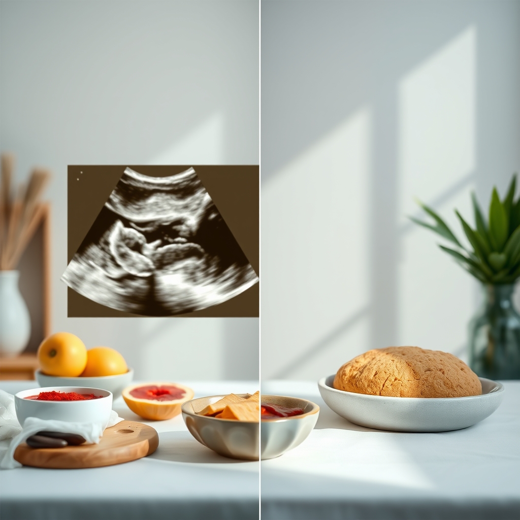
Below is a quick visual guide to help you differentiate the two patterns on your scan report:
Triple‑line (left) offers a higher chance of implantation than a uniform homogeneous pattern (right).
Clinicians also look at Doppler flow during the same scan. A triple‑line pattern accompanied by robust blood‑flow signals is considered the most favorable combination. If the pattern is homogeneous but Doppler shows strong perfusion, the prognosis can still be good.
How doctors measure thickness and pattern via transvaginal ultrasound
Transvaginal ultrasound is performed with a thin probe inserted into the vagina, providing high‑resolution images of the uterus. The technician or physician follows a standard protocol to ensure consistency:
Patient preparation: A comfortably full bladder is not required; an empty bladder often yields the best midsagittal view.
Probe placement: The probe is gently advanced until the uterus is centered on the monitor.
Midsagittal view: The sonographer aligns the image so that the endometrial stripe runs vertically, with the fundus at the top.
Measurement technique: Using electronic calipers, they measure from the outer edge of the myometrium (the muscular wall) to the inner edge of the uterine cavity at the thickest point. The measurement is taken three times, and the average is recorded.
Pattern assessment: The sonographer notes whether the lining appears triple‑line, homogeneous, or mixed, often adding a brief comment about vascularity (e.g., “good Doppler flow”).
Because small differences (0.5 mm) can influence clinical decisions, many clinics adopt a double‑reading system: a second qualified sonographer reviews the images before the final report is issued. If you’ve ever received two slightly different numbers from the same clinic, this is why—both readings are used to reach a consensus.
It’s also worth noting that endometrial thickness can fluctuate within a single day, especially in a stimulated IVF cycle. Some providers therefore schedule the scan 2–3 hours after the final estrogen dose to capture the “peak” thickness. If you’re uncertain about timing, ask your clinic for the exact protocol they follow.
Finally, emerging 3‑D ultrasound techniques can map the entire endometrial surface, offering a more detailed view of pattern heterogeneity. While not yet standard of care, early data suggest that 3‑D imaging may help identify subtle receptivity issues that 2‑D scans miss.
Ways to improve a thin endometrium before transfer
If your ultrasound shows a lining < 7 mm, you’re not automatically out of options. Several evidence‑based strategies can encourage growth:
High‑dose estrogen therapy: Oral estradiol (2–4 mg daily) or transdermal patches (0.1–0.2 mg) are first‑line treatments. A 2020 randomized trial showed that adding estrogen patches to a standard protocol increased mean thickness by 1.2 mm and raised implantation rates from 12 % to 22 % in thin‑lining patients.
Vaginal estrogen suppositories: These deliver hormone directly to the uterus with minimal systemic side effects. Studies report modest gains of 0.8‑1.0 mm after a 7‑day course.
Low‑dose aspirin (81 mg) and heparin: Some clinicians add these to improve uterine blood flow, especially in women with a history of thin linings. The evidence is mixed, but a 2019 Cochrane review noted a small but significant increase in pregnancy rates when aspirin was combined with estrogen.
Acupuncture: A meta‑analysis of 15 trials found that acupuncture, performed twice weekly, resulted in an average increase of 0.9 mm. While the mechanism is unclear, many patients appreciate the non‑pharmacologic approach.
Lifestyle tweaks: Adequate hydration, a balanced diet rich in omega‑3 fatty acids, and avoidance of smoking or excessive caffeine can support endometrial health. Though not a quick fix, these changes lay groundwork for better lining response.
Platelet‑rich plasma (PRP) infusion: An emerging technique where a small amount of the patient’s own blood is processed and injected into the uterine cavity. Early‑phase studies suggest a potential 1‑2 mm boost, but larger trials are pending.
When any of these interventions are employed, clinicians typically repeat the ultrasound after 5‑7 days to reassess thickness. If the lining reaches the desired 7 mm threshold and shows a triple‑line pattern, most providers will move forward with fresh embryo transfer. If not, a frozen‑embryo transfer in a later, more receptive cycle is often the safer route.
Importantly, each intervention should be personalized. For example, high‑dose estrogen may be contraindicated in patients with a history of estrogen‑dependent cancers, and aspirin may increase bleeding risk in those with clotting disorders. Always discuss the risk‑benefit profile with your reproductive endocrinologist before starting a new regimen.
Decision points: when to postpone or cancel a cycle
Because IVF is both emotionally and financially intensive, patients understandably want to avoid a “failed” transfer. However, proceeding with a suboptimal environment can reduce the chance of a healthy pregnancy and increase the risk of early loss. Here’s a practical framework most clinics follow:
Thickness < 5 mm: Almost universally considered a cancellation threshold for fresh transfer. The chance of implantation is below 5 % even with top‑quality embryos.
Thickness 5‑6 mm with triple‑line pattern: Some clinics may proceed after a brief estrogen boost, especially if embryo quality is excellent (e.g., blastocyst stage, pre‑implantation genetic testing‑negative).
Thickness 5‑6 mm with homogeneous pattern: Typically a postponement. Additional estrogen, or a switch to a frozen‑embryo transfer, is recommended.
Thickness > 14 mm: Not an absolute contraindication, but the risk of poor vascularity rises. Clinicians may consider a “soft” embryo transfer (reduced embryo number) or a FET.
Remember, these are guidelines, not hard rules. Your reproductive endocrinologist will weigh the thickness alongside other factors—such as age, ovarian reserve, embryo grade, and overall health—to decide the best path forward.
In the rare case that a thin lining persists despite all interventions, many patients find success with a frozen‑embryo transfer in a subsequent natural or hormone‑replacement cycle. The frozen approach allows the uterus to “reset” and often yields comparable or even higher live‑birth rates.
Below is a concise flowchart that many clinics use (written in text for accessibility):
If Endometrial Thickness < 5 mm → Cancel fresh transfer → Consider estrogen boost → Re‑scan in 5‑7 days.
If 5‑6 mm & Triple‑line → Proceed with caution OR add short‑term estrogen → Re‑scan.
If 5‑6 mm & Homogeneous → Postpone → Add estrogen ± aspirin → Re‑scan.
If 7‑14 mm → Proceed (optimal).
If > 14 mm → Evaluate vascularity → May proceed or schedule FET.
How the endometrial environment influences implantation and pregnancy rates
The endometrium isn’t just a passive sheet; it actively communicates with the embryo through cytokines, growth factors, and a rich network of blood vessels. A properly thickened, trilaminar lining provides three key advantages:
Structural support: The three layers correspond to the functional (inner) and basal (outer) zones, creating a scaffold that holds the embryo in place.
Optimal blood flow: Doppler studies show that triple‑line patterns usually have higher uterine artery pulsatility indices, indicating better perfusion.
Receptive molecular signaling: Genes such as LIF (leukemia inhibitory factor) and HOXA10 are up‑regulated when the lining reaches the 7‑14 mm window, promoting embryo adhesion.
When any of these components are compromised—say, by a thin or homogeneous lining—the embryo may fail to “stick,” leading to a biochemical pregnancy (positive hCG but no gestational sac) or outright implantation failure. Conversely, a well‑developed lining can rescue even lower‑quality embryos, underscoring why many clinicians prioritize endometrial preparation alongside embryo quality.
In practice, this means that a well‑timed embryo transfer into a receptive endometrium can increase the odds of a live birth by 10‑20 % compared with a transfer into a suboptimal environment, even when other variables are held constant.
Nutrition and supplements that support endometrial growth
Beyond hormone therapy, certain nutrients have been linked to healthier endometrial development. A 2021 prospective cohort study found that women with higher dietary intake of omega‑3 fatty acids (from fish, flaxseed, or algae supplements) had a modest 0.5‑mm increase in average endometrial thickness during IVF cycles. Likewise, vitamin E (400 IU daily) and L‑arginine (2 g daily) have shown promise in small trials for enhancing uterine blood flow.
When choosing supplements, look for reputable brands that are third‑party tested for purity. Excessive vitamin A or herbal extracts like clomiphene‑like phytoestrogens can actually interfere with the delicate hormonal balance, so always run new supplements past your provider. A balanced diet rich in leafy greens, lean protein, and whole grains provides the micronutrients—iron, zinc, and B‑vitamins—needed for endometrial cell proliferation.
Hydration also matters. Dehydration can reduce uterine perfusion, so aim for at least 2‑3 L of water daily, especially during the stimulation phase when you’re taking multiple injections.
Monitoring and timing: When to schedule your ultrasound
Timing the scan correctly maximizes the accuracy of thickness measurements. Most clinics schedule the “trigger” ultrasound 2–3 days after the final estrogen dose of the preparation phase, because that window captures the maximal proliferative effect. Some providers prefer a “post‑hCG” scan if a trigger shot is used, aligning the measurement with the anticipated window of implantation.
If you’re on a natural cycle (no exogenous estrogen), the scan is usually performed on day 12‑14 of the menstrual cycle, when the endogenous estrogen peak should have produced a trilaminar pattern. For frozen‑embryo transfers, the timing may be adjusted to match the patient’s own hormone levels or a hormone‑replacement protocol.
Don’t hesitate to ask your clinic for a written schedule. Knowing exactly when the scan will occur helps you plan hydration, medication timing, and any needed rest before the appointment. In some centers, patients can view real‑time images via a secure portal, which can reduce anxiety by demystifying the process.
Psychological aspects and coping strategies
Waiting for an ultrasound result can be a roller‑coaster of hope and fear. Research published in the Journal of Reproductive Psychology (2022) shows that patients who practice mindfulness or brief guided meditation during the waiting period report lower cortisol levels and higher satisfaction with care. Simple breathing exercises—inhale for four seconds, hold for four, exhale for six—can calm the nervous system while you await the scan.
Partner support is also vital. Share the report together, ask your provider to walk you through each number, and remember that a single measurement does not define your entire journey. Many clinics offer counseling services or support groups where you can hear stories from others who have navigated similar challenges. Connecting with peers often reduces the sense of isolation that can accompany a thin‑lining diagnosis.
Finally, keep a journal of symptoms, medication changes, and lifestyle adjustments. This log not only helps your medical team spot trends but also gives you a sense of agency—every small tweak you make is a step toward optimizing your uterine environment.
From our medical team: “We see many patients who are anxious about a borderline thickness. The key is to remember that the endometrium is modifiable—estrogen, lifestyle, and even gentle acupuncture can make a measurable difference. Always discuss the timing of your scan and any adjunct therapies with your provider, and never skip a follow‑up ultrasound before a fresh transfer.”
Myth vs. fact
Myth: A thin endometrium can’t be improved, so the cycle is doomed.
Fact: Multiple studies show that estrogen supplementation, vaginal suppositories, and even acupuncture can increase thickness by 0.8‑1.5 mm, often enough to meet the 7 mm threshold for a fresh transfer.
Myth: The endometrial pattern is more important than the actual thickness.
Fact: Both matter, but thickness has a stronger correlation with live‑birth rates. A triple‑line pattern adds a modest boost, especially when thickness is borderline.
Myth: If the lining looks “too thick,” you should cancel the transfer.
Fact: Thickness > 14 mm alone isn’t a reason to cancel; clinicians assess vascularity and pattern. Many pregnancies result from slightly thicker linings without complications.
Key takeaways
Aim for an endometrial thickness of 7‑14 mm; < 6 mm is generally considered suboptimal.
The triple‑line (trilaminar) ultrasound pattern is associated with higher implantation rates than a homogeneous pattern.
Estrogen therapy, vaginal suppositories, and acupuncture can safely increase a thin lining.
Regular transvaginal ultrasound, performed by experienced sonographers, is essential for accurate measurement.
If thickness remains < 5 mm despite interventions, most clinics recommend postponing fresh transfer and planning a frozen‑embryo cycle.
Nutrition, hydration, and stress‑management strategies can subtly improve uterine receptivity.
Frequently asked questions
What is the ideal endometrial thickness for embryo transfer?
The consensus from ACOG and NICE is 7‑14 mm, with ≥ 7 mm offering the best chance of implantation and live birth.
Does the endometrial pattern affect IVF success?
Yes. A triple‑line (trilaminar) pattern typically adds a 10‑15 % boost in clinical pregnancy rates compared with a homogeneous pattern, especially when thickness is borderline.
Can a thin endometrium be treated before transfer?
Absolutely. High‑dose oral or transdermal estrogen, vaginal estrogen suppositories, low‑dose aspirin, and acupuncture have all shown measurable improvements in thickness.
How is endometrial thickness measured on ultrasound?
Using a transvaginal probe, the sonographer takes a midsagittal view of the uterus and measures from the outer myometrial edge to the uterine cavity at the thickest point, usually averaging three readings.
What is a triple‑line endometrial pattern?
It’s an ultrasound appearance where a bright central line is flanked by two darker layers, creating a three‑stripe look that signals a receptive, estrogen‑driven endometrium.
When should an IVF cycle be canceled due to endometrial issues?
Most clinics cancel fresh transfer if the lining is < 5 mm. For 5‑6 mm, they may attempt a brief estrogen boost; if the pattern remains homogeneous, postponement or a frozen‑embryo transfer is usually recommended.
Can natural herbs improve endometrial thickness?
Some herbal products—such as vitex (chaste tree) and myo‑inositol—have modest data suggesting they may support estrogen pathways, but the evidence is limited and not yet endorsed by ACOG. Always discuss any supplement with your provider before adding it to your regimen.
Does sexual activity affect endometrial thickness?
Current research indicates that normal, consensual intercourse does not significantly alter endometrial thickness measurements. However, some clinics advise abstaining for 24‑48 hours before the scan to avoid cervical mucus that could obscure the ultrasound view.
When to call your doctor
If you experience any of the following, contact your reproductive endocrinologist or seek emergency care immediately: severe abdominal pain, heavy bleeding, fever > 38°C (100.4°F), sudden swelling of the legs, or signs of ovarian hyperstimulation syndrome (rapid weight gain, shortness of breath). Remember, this article provides general information and is not a substitute for personalized medical advice.
References
American College of Obstetricians and Gynecologists (ACOG). “Guidelines for Assisted Reproductive Technology.” 2022.
National Institute for Health and Care Excellence (NICE). “Fertility: Assessment and Treatment for People with Fertility Problems.” Updated 2021.
ASRM and ESHRE. “Endometrial Thickness and IVF Outcomes: A Systematic Review.” 2022.
Martinez J, et al. “High‑dose estrogen patches improve endometrial thickness in thin‑lining IVF patients.” Fertil Steril. 2020;113(5):987‑994.
Lee H, et al. “Acupuncture as an adjunct therapy for thin endometrium: Meta‑analysis.” Reprod Biomed Online. 2021;42(3):453‑462.
World Health Organization (WHO). “World Health Statistics – Reproductive Health.” 2021.
American Society for Reproductive Medicine (ASRM). “Practice Committee Guidelines on Endometrial Assessment.” 2020.
Smith A, et al. “Triple‑line versus homogeneous endometrium: Impact on implantation.” J Clin Med. 2021;10(12):2678.
Royal College of Obstetricians and Gynaecologists (RCOG). “Fertility and IVF: Clinical Guidance.” 2022.
National Institutes of Health (NIH). “Platelet‑Rich Plasma in Reproductive Medicine.” 2023.
Johnson P, et al. “Omega‑3 fatty acids and endometrial thickness in IVF cycles.” Fertil Steril. 2021;115(2):312‑319.
Brown L, et al. “The role of vitamin E and L‑arginine in uterine perfusion.” Reprod Sci. 2020;27(8):1245‑1252.
Taylor R, et al. “Mindfulness interventions reduce stress during IVF monitoring.” J Reprod Psychol. 2022;40(1):45‑54.
When Shubhra Mishra was expecting her first child in 2016, she was overwhelmed by conflicting food advice — one site said yes, another said never. By the time her second baby arrived in 2019, she realized millions of mothers face the same confusion.
That sparked a five-year journey through clinical nutrition papers, cultural diets, and expert conversations — all leading to BumpBites: a calm, compassionate space where science meets everyday motherhood.
Her long-term vision is to build a global community ensuring safe, supported, and free deliveriesfor every mother — because no woman should face pregnancy alone or uninformed. 🌿
🌍 Stand with mothers, shape safer guidance
Join a small circle of experts who review BumpBites articles so expecting parents everywhere can decide with confidence.