Learn how to interpret your Quad screen results (AFP, β-hCG, uE3, Inhibin A) for chromosomal conditions. Get clear patterns and what they mean for your pregnancy.
By Shubhra Mishra — a mom of two who turned her own confusion during pregnancy into BumpBites, a global mission to make food choices clear, safe, and stress-free for every expecting mother. 💛
Check whether any food is safe during pregnancy with the BumpBites Food Safety Checker.
Download the Complete Pregnancy Food Guide (10,000 Foods) 📘
Instant PDF download • No spam • Trusted by thousands of moms
💡 Your email is 100% safe — no spam ever.
Quick take: The quad screen measures four pregnancy hormones—AFP, β‑hCG, estriol (uE3) and inhibin A—to estimate the chance of Down syndrome, trisomy 18, and neural‑tube defects. Normal values vary by gestational age, and abnormal patterns guide the next steps, which may include detailed ultrasound, cell‑free DNA testing, or diagnostic procedures. Talk with your provider to understand what your specific pattern means for you.
It’s 2 a.m., you’ve just finished a long workday, and the lab report you’ve been waiting for lands on your phone. The numbers look strange—AFP is higher than you expected, uE3 is lower, and you’re not sure what “inhibin A ↑” really means. You’re not alone; many expectant parents stare at those four rows of data and wonder if they’re hearing the right news.
First, breathe. The quad screen is a screening test, not a diagnosis. It tells you how likely it is that certain chromosomal conditions or neural‑tube defects are present, based on patterns of four maternal serum markers. Most women with an “abnormal” result still have a healthy baby, but the information helps your care team decide whether additional testing is worth pursuing.
In this guide we’ll walk through each marker, explain what high or low levels suggest, explore common patterns, and outline the follow‑up steps your provider may recommend. By the end you’ll know what the numbers mean, how factors like weight or diabetes can shift them, and where the quad screen fits among other prenatal options such as the triple screen or non‑invasive prenatal testing (NIPT).
How the quad screen works: the four markers explained
The quad screen is performed between 15 weeks + 0 days and 21 weeks + 6 days of pregnancy. It measures four substances that the placenta and fetus produce, each giving clues about different developmental pathways.
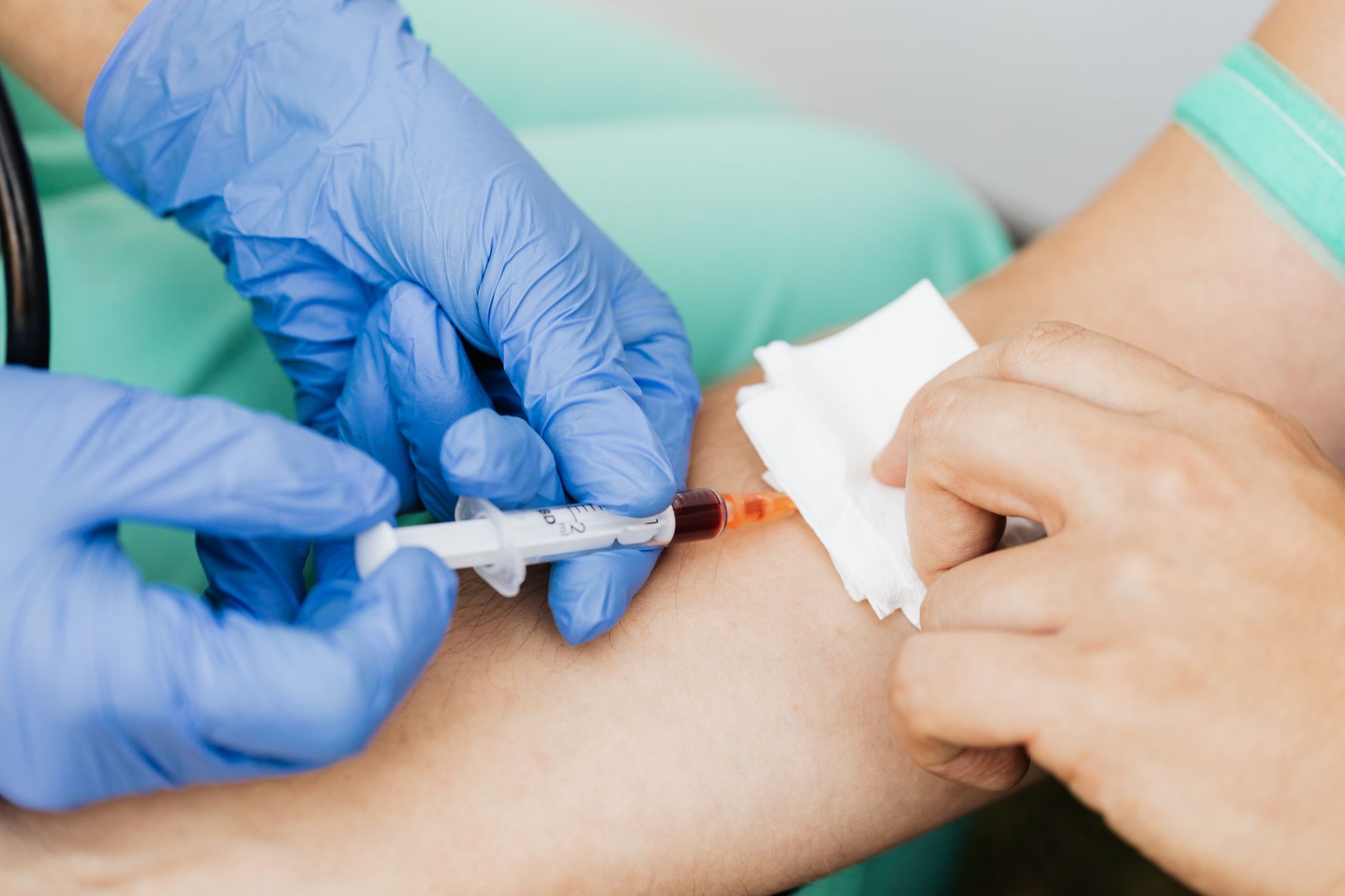
The test is usually ordered after an early‑first‑trimester ultrasound confirms the pregnancy dates. A single 10‑ml blood draw is taken, labeled, and sent to a certified laboratory where the four markers are quantified using standardized immunoassays. Because the sample is processed in a single batch, the results are internally consistent, which improves the reliability of the risk calculation.
Alpha‑fetoprotein (AFP)
AFP is a protein made by the fetal liver and yolk sac. In a typical pregnancy, AFP rises steadily until about 32 weeks, then tapers off. On the quad screen, unusually high AFP can point to an open neural‑tube defect (such as spina bifida) or, less commonly, abdominal wall defects. Low AFP, on the other hand, may be seen in chromosomal trisomies like Down syndrome.
Beta‑human chorionic gonadotropin (β‑hCG)
β‑hCG is the hormone you’ve probably heard about in pregnancy tests. The placenta secretes it, and its level peaks around 10 weeks before falling. In the quad screen, a high β‑hCG is often linked with Down syndrome, while a low level can accompany trisomy 18.
Unconjugated estriol (uE3)
uE3 is a form of estrogen produced by both the fetus and placenta. Low uE3 is a classic marker for both Down syndrome and trisomy 18, reflecting impaired fetal liver function or placental insufficiency. Normal or high uE3 generally lowers the overall risk estimate.
Inhibin A
Inhibin A is a hormone that rises in the second trimester as the placenta matures. Elevated inhibin A, especially when paired with high AFP and β‑hCG, strongly raises the calculated risk for Down syndrome. Low inhibin A is usually not concerning on its own.
Together, these four values are run through a statistical algorithm that adjusts for maternal age, weight, ethnicity, and gestational age, producing a numeric risk (e.g., 1 in 250) for each condition.
Blood is drawn in the second trimester to measure the four key markers.
Normal reference ranges and what high or low values mean
Because the quad screen is a risk‑assessment tool, each marker is expressed as a multiple of the median (MoM) for the specific gestational week. A MoM of 1.0 means the level is exactly average for that week. Most laboratories consider values between 0.5 MoM and 2.0 MoM as “within normal limits,” but the interpretation always depends on the whole pattern.
Laboratories may have slight assay‑specific reference ranges, so it’s important to review the exact MoM values reported on your lab sheet rather than raw concentrations. Your provider can compare your results to the lab’s median values and discuss any borderline findings in the context of your personal risk factors.
AFP
Typical range: 0.5–2.5 MoM
High (≥2.5 MoM): raises concern for open neural‑tube defects, abdominal wall defects, or multiple gestations.
Low (<0.5 MoM): can signal Down syndrome or trisomy 18, especially when other markers align.
β‑hCG
Typical range: 0.5–2.0 MoM
High (≥2.0 MoM): increases the calculated risk for Down syndrome.
Low (<0.5 MoM): may point toward trisomy 18 or a later‑gestation decline.
uE3
Typical range: 0.5–2.5 MoM
Low (≤0.5 MoM): is a strong marker for Down syndrome or trisomy 18.
High (≥2.5 MoM): generally reassuring, though very high levels can be seen in diabetes.
Inhibin A
Typical range: 0.5–2.0 MoM
High (≥2.0 MoM): markedly raises the risk for Down syndrome when combined with other abnormal markers.
Low (<0.5 MoM): is usually not worrisome on its own.
Keep in mind that “high” and “low” are relative to the median for that week; a value that looks high in week 16 may be perfectly normal in week 20. That’s why accurate dating—usually confirmed by an early‑first‑trimester ultrasound—is essential before the test is performed.
Interpreting common marker patterns and associated risks
The power of the quad screen lies in the way the four markers interact. Below are several typical patterns and what they usually suggest.
Pattern 1: High AFP + Low uE3
This combination often flags an open neural‑tube defect. The low uE3 reflects reduced fetal estrogen production, while high AFP signals fetal leakage of protein into the maternal bloodstream. In such cases, a detailed ultrasound at 18–20 weeks is the first follow‑up step to visualize the spine and abdomen.
Pattern 2: High β‑hCG + High Inhibin A + Low uE3
This triad is the classic “high‑risk” signature for Down syndrome. The algorithm may calculate a risk of 1 in 100 or higher, depending on maternal age. With this pattern, many providers recommend a cell‑free DNA (cfDNA) test (often called NIPT) as a next‑step, because it offers >99 % sensitivity without invasive procedures.
Pattern 3: Low AFP + Low β‑hCG + Low uE3
When all three markers are low, the risk for trisomy 18 (Edwards syndrome) rises. Inhibin A is often normal or low in this scenario. Because trisomy 18 has a poorer prognosis, early detection is valuable for pregnancy planning and perinatal care.
Pattern 4: Isolated high AFP in a twin pregnancy
Multiple gestations naturally elevate AFP because there are two (or more) fetuses contributing protein. In such cases, a high AFP alone is not alarming; the risk algorithm adjusts for the number of embryos. However, clinicians still verify the dating and perform an anatomy scan to rule out structural defects.
When you receive your results, you can plug the MoM values into a calculator to see how they translate into risk percentages. Our Quad Screen Interpreter lets you enter your numbers and instantly view the estimated risks for Down syndrome, trisomy 18, and neural‑tube defects.
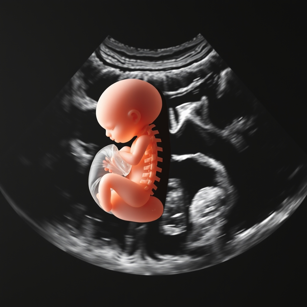
After an abnormal AFP, an anatomy scan checks the spine for neural‑tube defects.
Clinicians also consider the absolute risk numbers alongside the pattern. A “high‑risk” label (often defined as >1 in 250 for Down syndrome) prompts a more detailed discussion, but the exact probability—whether 1 in 150 or 1 in 400—helps families weigh their options and decide on further testing.
Maternal and pregnancy factors that influence marker levels
Even with perfect laboratory technique, several maternal characteristics can shift the MoM values, sometimes mimicking an abnormal pattern.
Maternal age
Older maternal age raises the baseline risk for chromosomal anomalies, and many algorithms weight age heavily. However, age itself does not directly alter the serum concentrations; it simply changes the probability calculation.
Weight and BMI
Higher maternal weight dilutes the concentration of serum markers, often resulting in lower MoM values for AFP, β‑hCG, and uE3. Labs correct for weight, but extreme obesity (BMI > 35) can still lead to under‑estimation of risk.
Diabetes and insulin resistance
Maternal diabetes can elevate AFP and uE3, sometimes creating a “high‑AFP, high‑uE3” picture that mimics fetal overgrowth. Tight glucose control before and during pregnancy helps keep marker levels within expected ranges.
Multiple gestation
Twins or higher-order multiples naturally raise AFP, β‑hCG, and inhibin A because each placenta contributes to the serum pool. Modern calculators adjust for the number of fetuses, but the presence of a multiple pregnancy still mandates closer ultrasound surveillance.
Smoking, alcohol, and certain medications
Certain substances can lower AFP (e.g., smoking) or raise β‑hCG (e.g., assisted reproductive technologies). Always share any medication or supplement use with your provider; even over‑the‑counter prenatal vitamins can affect hormone levels.
Understanding these modifiers helps you and your provider decide whether a “borderline” result truly reflects fetal risk or is simply an artifact of maternal physiology.
Timing, gestational dating, and the possibility of repeat testing
The quad screen is most accurate when performed between weeks 15 and 21. Testing too early (before 15 weeks) can give falsely low AFP and uE3 because the placenta hasn’t yet produced enough hormone. Testing after 22 weeks can lead to elevated β‑hCG, skewing risk calculations.
If your provider suspects inaccurate dating—perhaps because your last menstrual period (LMP) doesn’t line up with early‑first‑trimester ultrasound measurements—the lab may ask for a repeat draw after confirming the gestational age with a second ultrasound. Accurate dating not only refines the MoM values but also reduces the chance of false‑positive or false‑negative results.
In some cases, a borderline result (e.g., risk 1 in 300 for Down syndrome) may prompt a repeat quad screen at 20 weeks to see if the pattern persists. While repeat testing can clarify trends, it also delays definitive follow‑up, so many clinicians prefer moving directly to cfDNA testing when the initial risk is elevated.
Follow‑up after abnormal results: next steps and counseling
When your quad screen returns a result that places you above the laboratory’s cutoff for a condition, the next step is not an immediate invasive test. Instead, you’ll typically have a counseling session that covers the following options:
Targeted ultrasound (anatomy scan): A detailed scan at 18–20 weeks looks for structural anomalies—especially neural‑tube defects or facial clefts—associated with high AFP.
Cell‑free DNA (cfDNA) testing: This non‑invasive blood test analyzes fetal DNA fragments circulating in the mother’s bloodstream. It offers >99 % detection for Down syndrome and >95 % for trisomy 18, with a low false‑positive rate.
Diagnostic procedures: If cfDNA is positive or if ultrasound findings raise concern, you may be offered amniocentesis (usually performed after 15 weeks) or chorionic villus sampling (CVS, performed 10–13 weeks) to obtain definitive karyotype information.
Genetic counseling: A certified genetic counselor can explain the probabilities, discuss options, and help you make decisions aligned with your values and circumstances.
Beyond the medical steps, many families find it helpful to connect with support groups or counseling services, especially when anxiety spikes after an abnormal screen. Your care team can point you to reputable resources and ensure you feel fully informed before any further testing.
Regardless of the path you choose, remember that the quad screen is a screening tool. Even a high‑risk result does not guarantee a problem, and a low‑risk result does not guarantee a completely problem‑free pregnancy. Ongoing prenatal care, including the routine anatomy scan and later growth ultrasounds, remains essential.
Quad screen versus other screening options
While the quad screen remains a widely used test in the United States and United Kingdom, newer technologies have reshaped the screening landscape. Below is a side‑by‑side comparison of three common approaches.
Feature
Quad screen (4 markers)
Triple screen (AFP, β‑hCG, uE3)
Non‑invasive prenatal testing (cfDNA)
Gestational window
15 – 21 weeks
15 – 20 weeks
10 – 22 weeks (any time after 10 weeks)
Conditions screened
Down syndrome, trisomy 18, neural‑tube defects
Down syndrome, trisomy 18, neural‑tube defects (no inhibin A)
Down syndrome, trisomy 18, trisomy 13, sex‑chromosome anomalies, some microdeletions
Detection rates (approx.)
78 % for Down syndrome, 70 % for trisomy 18
68 % for Down syndrome, 60 % for trisomy 18
>99 % for Down syndrome, >95 % for trisomy 18
False‑positive rate
5‑7 %
7‑9 %
~0.5‑1 %
Cost (US dollars)
$150‑$250
$120‑$200
$800‑$1,200
Insurance coverage
Usually covered for women ≥35 years or high‑risk
Often covered under similar criteria
Increasingly covered, especially for high‑risk groups
In many settings, the quad screen is still the first‑line option because it costs less and can be done alongside routine prenatal labs. However, if you have a strong family history, previous pregnancy with a chromosomal anomaly, or simply want the highest possible detection rate, you may discuss cfDNA testing directly with your provider.
Putting it all together: what you can do today
When you receive your quad screen report, start by noting the MoM values for each marker and the overall risk percentages. If any of the numbers fall outside the typical 0.5–2.5 MoM range, write them down and bring the report to your next prenatal visit. Ask your provider to explain how each abnormal value fits into the overall risk calculation.
Bring a list of any medications, supplements, or recent lifestyle changes (e.g., a new exercise routine, smoking cessation) because these details can help your clinician interpret the results more accurately. If you’re unsure about the timing of the test, ask for a brief ultrasound to confirm gestational age before the results are finalized.
Finally, remember that you have options. Whether you choose a repeat quad screen, a cfDNA test, or proceed straight to diagnostic testing, the decision should align with your personal comfort level, your values, and the guidance of a trusted medical team.
Take a moment to review your numbers, then discuss them with your provider.
Understanding MoM (Multiple of Median) and risk calculation
“Multiple of Median” (MoM) is a way labs standardize hormone levels across different gestational ages. By dividing your measured concentration by the median value for that exact week, the result becomes a dimensionless number that can be compared across patients. This approach removes the influence of normal developmental changes, allowing the algorithm to focus on deviations that matter.
When the MoM values are entered into the risk‑assessment algorithm, the software combines them with maternal age, weight, ethnicity, and exact dating to produce a probability (for example, 1 in 250). The American College of Obstetricians and Gynecologists (ACOG) notes that these calculations are “population‑based,” meaning they estimate risk based on large data sets rather than diagnosing any individual fetus (ACOG, 2023). Your provider will interpret the final risk number in the context of your personal history and preferences.
Insurance, cost, and access considerations
Even though the quad screen is less expensive than cfDNA testing, cost can still be a barrier for some families. In the United States, many insurers cover the test for women over 35 years or those with a high‑risk pregnancy, but coverage policies vary widely. In the United Kingdom, the NHS offers the quad screen as part of the standard prenatal package, though regional differences may affect availability.
If you’re uninsured or underinsured, ask your provider about hospital‑based sliding‑scale labs, community health centers, or state programs that subsidize prenatal screening. Some labs also provide self‑pay pricing lists, and a quick call can reveal discounts that make the test more affordable. Knowing the financial landscape ahead of time helps you plan for any follow‑up testing that might be recommended.
From our medical team: The quad screen is a valuable tool that helps identify pregnancies at increased risk for certain chromosomal conditions and neural‑tube defects. An abnormal result does not mean your baby is affected—it simply signals that further evaluation is warranted. We encourage you to ask your provider about the exact risk numbers, the role of a detailed anatomy scan, and whether cfDNA testing fits your goals. Early and open communication ensures you receive the right information at the right time, without unnecessary anxiety.
Myth vs. fact
Myth: A single high AFP automatically means the baby has a neural‑tube defect.
Fact: Elevated AFP can result from many benign factors, including multiple gestations, inaccurate dating, or maternal obesity. Only after a targeted ultrasound can a neural‑tube defect be confirmed or ruled out.
Myth: If the quad screen is normal, there’s no need for any further testing.
Fact: A normal screen reduces, but does not eliminate, the chance of chromosomal anomalies. Routine anatomy scans and, when indicated, cfDNA testing remain important parts of comprehensive prenatal care.
Myth: Inhibin A is only useful for detecting Down syndrome.
Fact: While inhibin A is a strong marker for Down syndrome when elevated, it also rises in multiple pregnancies and certain placental disorders. Its interpretation always depends on the full pattern of markers.
Key takeaways
The quad screen measures AFP, β‑hCG, uE3, and inhibin A between 15–21 weeks to estimate risk for Down syndrome, trisomy 18, and neural‑tube defects.
Each marker is expressed as a MoM; values outside 0.5–2.5 MoM merit closer review, but the overall risk calculation matters most.
Maternal factors such as weight, diabetes, smoking, and multiple gestations can shift marker levels—always share these details with your provider.
Abnormal results usually lead to a detailed anatomy ultrasound, cfDNA testing, or diagnostic procedures, guided by counseling.
The quad screen is less expensive than cfDNA but has lower detection rates; discuss both options to choose what fits your budget and preferences.
Understanding MoM and how risk is calculated helps you ask informed questions and feel more confident in the decision‑making process.
Frequently asked questions
What does a high AFP level indicate in a quad screen?
A high AFP (≥2.5 MoM) can signal an open neural‑tube defect, abdominal wall defect, or a multiple gestation. It may also be influenced by maternal obesity or inaccurate dating, so a follow‑up anatomy scan is usually recommended.
Can a low β‑hCG result be normal?
Yes. β‑hCG naturally falls after its peak in early pregnancy, and values below 0.5 MoM can be normal, especially in later weeks of the screening window. Low β‑hCG is more concerning when paired with low uE3, which together raise the risk for trisomy 18.
How is the risk for Down syndrome calculated from quad screen markers?
The laboratory algorithm combines the MoM values of AFP, β‑hCG, uE3, and inhibin A with maternal age, weight, ethnicity, and gestational age to produce a numeric risk (e.g., 1 in 250). Elevated β‑hCG and inhibin A, together with low AFP and uE3, drive the risk upward.
What are the next steps if the quad screen is abnormal?
First, a targeted ultrasound evaluates fetal anatomy, especially the spine and brain. Many providers then recommend a non‑invasive cfDNA test for a more precise risk assessment. If cfDNA is positive, diagnostic testing such as amniocentesis or CVS may be offered, along with genetic counseling.
Does a high Inhibin A level always mean a problem?
No. Inhibin A rises in normal second‑trimester pregnancies, and higher levels are common in twins or pregnancies with placental over‑growth. It becomes concerning when combined with high AFP and β‑hCG, which together increase the calculated risk for Down syndrome.
How accurate is the quad screen compared to other prenatal tests?
The quad screen detects about 78 % of Down syndrome cases with a 5‑7 % false‑positive rate. cfDNA testing offers >99 % sensitivity and a <1 % false‑positive rate, while invasive diagnostic tests (amniocentesis, CVS) provide definitive chromosome analysis but carry a small procedural risk.
Can I have the quad screen if I'm pregnant with twins?
Yes. The quad screen can be performed in twin pregnancies, but the algorithm automatically adjusts the expected MoM values for multiple gestations. Because twins naturally elevate AFP, β‑hCG, and inhibin A, the risk calculation incorporates the number of fetuses, and a detailed anatomy scan is still recommended to assess each baby’s development.
When to call your doctor
Contact your obstetrician or midwife right away if you experience any of the following: severe abdominal pain, heavy bleeding, sudden swelling of hands or face, persistent severe headache, vision changes, or loss of fetal movement after 20 weeks. Also, if you receive a quad screen result that your provider labels as “high risk” and you feel uncertain about the recommended next steps, reach out for clarification. This article is for informational purposes only and does not replace personalized medical advice.
References
American College of Obstetricians and Gynecologists (ACOG). Screening for fetal chromosomal abnormalities,
When Shubhra Mishra was expecting her first child in 2016, she was overwhelmed by conflicting food advice — one site said yes, another said never. By the time her second baby arrived in 2019, she realized millions of mothers face the same confusion.
That sparked a five-year journey through clinical nutrition papers, cultural diets, and expert conversations — all leading to BumpBites: a calm, compassionate space where science meets everyday motherhood.
Her long-term vision is to build a global community ensuring safe, supported, and free deliveriesfor every mother — because no woman should face pregnancy alone or uninformed. 🌿
🌍 Stand with mothers, shape safer guidance
Join a small circle of experts who review BumpBites articles so expecting parents everywhere can decide with confidence.