At your 7-week pregnancy sonography, you'll typically see the gestational sac, yolk sac, and a fetal pole with a flickering heartbeat. Learn what to expect during this exciting early scan.
By Shubhra Mishra — a mom of two who turned her own confusion during pregnancy into BumpBites, a global mission to make food choices clear, safe, and stress-free for every expecting mother. 💛
Check whether any food is safe during pregnancy with the BumpBites Food Safety Checker.
Quick take: Your 7-week pregnancy sonography is often your first glimpse into your pregnancy, confirming its location and viability. At this stage, you'll typically see the gestational sac, yolk sac, and a tiny fetal pole, often with a visible flickering heartbeat. It's a crucial scan for accurate dating and setting expectations for your pregnancy journey.
It's 7 weeks into your pregnancy, and you might be feeling a swirl of emotions – excitement, nervousness, and perhaps a little anxiety about what's happening inside. Your first ultrasound, often scheduled around this time, can feel like a huge milestone. You're probably wondering what exactly you'll see, what's normal, and what it all means. Take a deep breath; we're here to walk you through your 7-week pregnancy sonography, explaining everything you can expect in clear, reassuring terms.
This early scan is more than just a picture; it's a vital tool for your healthcare provider to gather important information about your pregnancy. It helps confirm your due date, check for viability, and ensure everything is progressing as it should. We understand that waiting for and going through this scan can be nerve-wracking, especially if you're feeling nauseous or experiencing other early pregnancy symptoms. Our goal is to empower you with knowledge, so you feel confident and prepared for what's ahead.
We'll cover everything from what structures you'll typically see, like the gestational sac and fetal pole, to understanding normal measurements and what to do if the scan doesn't show exactly what you expect. Remember, every pregnancy is unique, and your medical team is your best resource for personalized advice, but this guide will help you understand the basics of this exciting early peek at your baby.
Your 7-week ultrasound might be transvaginal for clearer early views.
What to Expect on Your 7-Week Ultrasound Scan?
Your 7-week ultrasound, or sonography, is often your first official introduction to your little one. It's an exciting, sometimes overwhelming, experience. The primary goal of this early scan is to confirm pregnancy, determine its location (to rule out ectopic pregnancy), establish an accurate due date, and assess the embryo's viability.
When you arrive for your appointment, you'll likely be asked to empty your bladder for a transvaginal ultrasound, which is very common at this early stage. This type of ultrasound involves inserting a small, lubricated probe into the vagina. While it might sound intimidating, most women find it only mildly uncomfortable, much like a pelvic exam. The transvaginal approach provides a much clearer and more detailed view of your uterus and embryo compared to a transabdominal (over the belly) ultrasound, especially when the embryo is still so tiny.
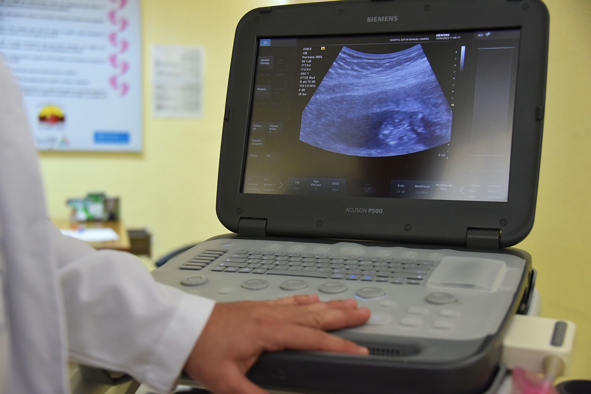
The sonographer, a trained medical professional who performs the scan, will explain what they're doing and what you're seeing on the screen. The image on the monitor might look a bit blurry or abstract to you at first, but they will point out key structures. Many parents tell us that seeing that first flicker of a heartbeat, even if it's just a tiny pulse on a black-and-white screen, is an incredibly emotional and unforgettable moment. One reader shared, "I was so nervous, my hands were shaking. But the moment the sonographer said 'there's the heartbeat,' a wave of relief washed over me. It was just a little blob, but it was MY blob, and it was real."
During the scan, the sonographer will be taking measurements and documenting what they see. This information is then reviewed by your doctor or midwife, who will discuss the findings with you. Don't hesitate to ask questions during or after the scan – it's your pregnancy, and you have every right to understand what's happening.
Can You See a Heartbeat at 7 Weeks Pregnant?
Yes, in most cases, you absolutely can see a heartbeat at 7 weeks pregnant! This is often the most anticipated and reassuring part of an early ultrasound. The tiny flicker you'll see on the screen represents the earliest cardiac activity of your embryo, and its presence is a strong indicator of a viable pregnancy.
At 7 weeks gestation, the heart has usually developed enough to begin its rhythmic contractions. The sonographer will typically measure the heart rate, which is an important indicator of healthy development. A normal heart rate at this stage can vary, but generally falls within the range of 110 to 160 beats per minute (bpm). Some medical guidelines suggest a range of 100-120 bpm at 6-7 weeks, increasing as the embryo develops further. Don't be alarmed if it's on the lower end of this range; sometimes, if your dates are slightly off and you're closer to 6 weeks, the heart rate might be a bit slower.
Seeing a heartbeat brings immense relief to many expectant parents. It provides tangible evidence that your pregnancy is progressing. However, it's also important to understand that if your dates are slightly off and you're not quite 7 full weeks pregnant, it's possible the heartbeat might not yet be visible. In such cases, your healthcare provider will likely recommend a follow-up scan in a week or two to check for development. This doesn't necessarily mean there's a problem; it simply means it might be a bit too early for a definitive finding.
The British Medical Journal (BMJ) highlights that the presence of a fetal heartbeat is the most reliable sign of viability in early pregnancy. If a heartbeat is detected and the embryo measurements are consistent with 7 weeks, it significantly reduces the risk of miscarriage moving forward. Our medical team often emphasizes that while the heartbeat is a wonderful sign, it's just one piece of the puzzle, and a comprehensive assessment of all structures is important.
What Should the Fetal Pole and Gestational Sac Look Like at 7 Weeks?
At 7 weeks, your ultrasound will reveal several key structures that confirm your pregnancy's progress. These include the gestational sac, the yolk sac, and the fetal pole. Understanding what each of these looks like and their significance can help you interpret your scan results.
The gestational sac is the first visible sign of pregnancy on an ultrasound, typically appearing around 4.5 to 5 weeks. At 7 weeks, it should be clearly visible within your uterus, appearing as a round or oval-shaped fluid-filled structure with a bright, reflective border. This sac is essentially the "home" for your developing embryo and contains amniotic fluid. Its size is measured to help confirm gestational age. The American College of Obstetricians and Gynecologists (ACOG) states that a gestational sac of at least 25 mm (about 1 inch) without a visible embryo is a sign of a failed pregnancy, but at 7 weeks, you should certainly see more than just the sac.
Inside the gestational sac, you'll see the yolk sac. This is another important structure, appearing as a small, round, fluid-filled sac, usually with a bright white ring around it. The yolk sac provides nourishment to the embryo until the placenta takes over, which happens later in the first trimester. It also plays a role in the development of the circulatory system. Its presence and normal appearance at 7 weeks are good indicators of a healthy, progressing pregnancy. The yolk sac usually measures between 3-6 mm at this stage.
Finally, the fetal pole is the earliest visual evidence of the embryo itself. At 7 weeks, the fetal pole is a tiny, elongated, C-shaped structure nestled against the yolk sac. It's often described as looking like a small "rice grain" or "bean sprout." This is the developing embryo, and it's where the heart activity will be detected. The fetal pole is measured from one end to the other, known as the Crown-Rump Length (CRL), which is crucial for accurate dating. The presence of a fetal pole with a visible heartbeat at 7 weeks is a very positive sign.
If you only see a gestational sac and yolk sac but no fetal pole, especially if the gestational sac is larger than expected for 7 weeks, your provider might suggest a follow-up scan. This could indicate that your dates are off, or it could be a sign of a blighted ovum, where a gestational sac develops but an embryo does not. Your medical team will guide you through any such findings with care and clarity.
The fetal pole is the earliest visible sign of your embryo.
What Are Normal Measurements for a 7-Week Fetus?
Understanding the measurements taken during your 7-week ultrasound can help you know what to expect and what's considered typical for this stage of development. The most important measurement taken at 7 weeks is the Crown-Rump Length (CRL), which measures the embryo from the top of its head (crown) to its bottom (rump).
At 7 weeks gestation, a healthy embryo typically has a CRL of approximately 9 to 13 mm (0.9 to 1.3 cm), which is roughly the size of a blueberry or a small raspberry. This measurement is incredibly important because it's the most accurate way to date your pregnancy in the first trimester. It helps pinpoint your estimated due date (EDD) with a margin of error of about +/- 3 to 5 days. This early dating is often more accurate than dating based on your last menstrual period (LMP), especially if you have irregular cycles.
In addition to the CRL, the sonographer will also measure the gestational sac diameter (GSD). At 7 weeks, a normal GSD typically ranges from 18 to 24 mm. As mentioned, the yolk sac is also measured, usually falling between 3 to 6 mm. All these measurements work together to provide a comprehensive picture of your pregnancy's progression.
Here's a table summarizing typical measurements at 7 weeks:
Structure
Typical Measurement at 7 Weeks
Significance
Crown-Rump Length (CRL)
9 - 13 mm (0.9 - 1.3 cm)
Most accurate for dating pregnancy; indicates embryo growth.
Gestational Sac Diameter (GSD)
18 - 24 mm
Confirms presence of pregnancy; should contain yolk sac and fetal pole.
Yolk Sac Diameter
3 - 6 mm
Provides early nourishment; indicates healthy development.
Heart Rate (FHR)
110 - 160 bpm
Key indicator of viability.
It's crucial to remember that these are averages. A measurement slightly outside this range doesn't automatically mean there's a problem. Small variations can occur due to slight differences in gestational age or individual growth patterns. Your healthcare provider will interpret all the findings in context and discuss any concerns with you. They might suggest a follow-up scan to monitor growth if measurements are borderline or if there's any uncertainty.
Is It Too Early for a 7-Week Ultrasound, or Is It Recommended?
For many expecting parents, the question often arises: is 7 weeks too early for an ultrasound? While some healthcare providers may wait until 8 or even 10 weeks, a 7-week ultrasound is widely recommended and often the standard practice, especially if there's uncertainty about dates, a history of miscarriage, or concerns about an ectopic pregnancy. It's not too early, and in fact, it offers significant benefits.
One of the primary reasons for a 7-week scan is to accurately date your pregnancy. As we discussed, the CRL measurement at this stage is highly precise, helping to establish your due date with remarkable accuracy. This is particularly useful for women with irregular menstrual cycles or those who aren't sure of their last menstrual period. Accurate dating is vital for future pregnancy management, including timing of screenings and interventions.
Another critical purpose of this early scan is to confirm the viability of the pregnancy. Seeing a fetal pole with a heartbeat at 7 weeks is a strong positive indicator that the pregnancy is progressing normally and significantly reduces the risk of early miscarriage. The Royal College of Obstetricians and Gynaecologists (RCOG) guidelines emphasize the importance of early viability scans in managing patient anxiety and providing reassurance.
Furthermore, a 7-week ultrasound can rule out an ectopic pregnancy, a serious condition where the embryo implants outside the uterus, most commonly in a fallopian tube. Detecting an ectopic pregnancy early is crucial for the mother's health. It can also identify multiple pregnancies (twins, triplets, etc.), which allows for early planning and specialized care. While a 7-week scan won't confirm the gender of the baby (it's far too early for that!), it provides foundational information about the pregnancy itself.
While the scan is highly beneficial, we understand that some people worry about the frequency of ultrasounds. According to major health organizations like the National Institute for Health and Care Excellence (NICE) in the UK, routine ultrasounds are considered safe and there's no evidence of harm to the developing baby from diagnostic ultrasound. It uses sound waves, not radiation, to create images.
What If Nothing Is Seen on My 7-Week Ultrasound?
It's natural to feel anxious when waiting for your first ultrasound, and one of the biggest worries is what happens if the scan doesn't show what's expected. If your 7-week ultrasound reveals "nothing" – meaning no fetal pole, no heartbeat, or perhaps only an empty gestational sac – it can be incredibly distressing. Take a deep breath; while this can be concerning, it doesn't always mean the worst. There are several reasons why this might happen, and your healthcare provider will guide you through the next steps with care.
The most common reason for not seeing expected structures is that your dates might be off. If you ovulated later than you thought, or if implantation occurred later, you might not be as far along as 7 weeks. For example, if you're truly only 5 or 6 weeks pregnant, it's very possible that a heartbeat or even a clear fetal pole might not yet be visible. In this scenario, your doctor will almost certainly recommend a follow-up ultrasound in 7-10 days to see if there's been development. Often, a subsequent scan reveals a healthy, growing embryo.
However, it's also important to be aware of other possibilities. If the gestational sac is larger than 25 mm and still empty, or if there's a fetal pole larger than 7 mm without a heartbeat, these are criteria often used to diagnose a miscarriage (sometimes referred to as a "missed miscarriage" or "blighted ovum" if there's a sac but no embryo). In a blighted ovum, the gestational sac forms and grows, but the embryo either never develops or stops developing very early on. You might not have experienced any typical miscarriage symptoms like bleeding or cramping, which is why it's called "missed."
Another rare but serious possibility is an ectopic pregnancy, where the pregnancy is located outside the uterus. While a 7-week scan aims to rule this out, if nothing is seen inside the uterus and you're experiencing symptoms like severe abdominal pain or bleeding, your doctor will investigate further with blood tests (hCG levels) and additional scans to locate the pregnancy. The Mayo Clinic emphasizes that a combination of ultrasound findings and hCG levels helps to diagnose these complex situations.
If your scan is inconclusive, your medical team will typically order serial blood tests to measure your human chorionic gonadotropin (hCG) levels. hCG is the "pregnancy hormone" that should rise predictably in early pregnancy. The rate of rise, combined with ultrasound findings, helps your doctor understand if the pregnancy is progressing normally. It's a waiting game that can be agonizing, but remember that your medical team is there to support you with information and emotional care, every step of the way.
If your scan is inconclusive, your medical team will provide clear next steps and support.
What Does a 7-Week Ultrasound Image Actually Show?
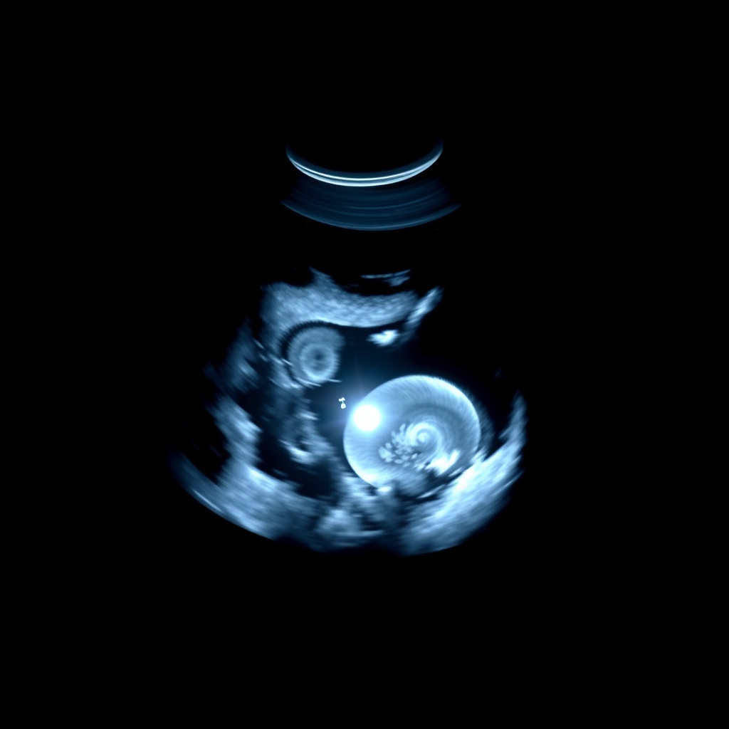
When you look at the ultrasound screen during your 7-week scan, it might initially look like a confusing grayscale image of blobs and shadows. However, once the sonographer or your doctor points out the key features, the image will start to make more sense and become incredibly meaningful. It's a window into the earliest stages of life!
The most prominent feature you'll see is the gestational sac. This will appear as a dark, fluid-filled circle or oval (the amniotic fluid) surrounded by a bright, reflective white ring. This white ring is the chorionic villi, which will eventually develop into part of the placenta, and it outlines the sac within your uterus. Imagine a tiny black bubble floating inside a larger, slightly darker space (your uterus).
Inside this gestational sac, you'll then be able to spot the yolk sac. This appears as a smaller, perfectly round, bright white ring, sometimes with a tiny black center (fluid) within it. It's usually nestled against the wall of the gestational sac. The yolk sac is typically very distinct and helps orient the sonographer to where the embryo should be.
Next to the yolk sac, you'll see the fetal pole. This is the embryo itself, and at 7 weeks, it will appear as a tiny, slightly elongated, C-shaped white structure, often described as resembling a grain of rice or a small kidney bean. It's very small, so you might need the sonographer to zoom in or point it out clearly. The most exciting part of seeing the fetal pole is the tiny, rapid, rhythmic flicker within or near it – this is the heartbeat. It doesn't look like a beating heart in the traditional sense, but rather a quick, repetitive flash or pulse on the screen.
The surrounding area will be your uterus, which will appear as a darker, muscular organ. You might also see parts of your ovaries, though they aren't the primary focus of this scan. The images are typically in black and white because ultrasound technology uses sound waves, not light, to create echoes that are then translated into these grayscale images. The different shades of gray represent varying densities of tissue, with fluid appearing dark and denser structures (like the chorionic villi or early embryo) appearing brighter. It's a truly amazing process that allows us to see such tiny details so early on.
What is the Purpose of a 7-Week Pregnancy Sonography?
A 7-week pregnancy sonography serves multiple crucial purposes beyond just giving you a first peek at your baby. It's an indispensable diagnostic tool that provides essential information for the safe and healthy management of your pregnancy. Understanding its full purpose can help you appreciate the importance of this early appointment.
One of the primary purposes, as highlighted by the American College of Obstetricians and Gynecologists (ACOG), is to confirm an intrauterine pregnancy. This means ensuring the pregnancy is located within your uterus, ruling out potentially life-threatening conditions like an ectopic pregnancy (where the fertilized egg implants outside the uterus, most commonly in a fallopian tube). If an ectopic pregnancy is suspected, an early ultrasound is vital for prompt diagnosis and management.
Another key objective is accurate gestational dating. By measuring the Crown-Rump Length (CRL) of the embryo, the sonographer can calculate your estimated due date (EDD) with remarkable precision in the first trimester. This early dating is more accurate than dating based on your last menstrual period, especially if your cycles are irregular or you're unsure of your ovulation date. An accurate due date is critical for scheduling future prenatal tests, monitoring fetal growth, and making decisions about labor and delivery.
The scan also helps to assess pregnancy viability. The presence of a fetal pole with a visible heartbeat at 7 weeks is a strong indicator of a viable, healthy pregnancy. While it doesn't guarantee everything will be smooth sailing, it significantly reduces the risk of early miscarriage. Conversely, if no heartbeat or fetal pole is seen when expected, it prompts further investigation and guidance from your provider.
Additionally, a 7-week ultrasound can detect multiple pregnancies. If you're carrying twins or more, this scan will typically identify separate gestational sacs or fetal poles, allowing for early specialized care tailored to multi-fetal pregnancies. It can also help to identify potential issues like molar pregnancy, a rare complication where abnormal tissue grows in the uterus instead of a healthy embryo.
In essence, the 7-week sonography provides foundational information that informs your entire prenatal care plan. It offers reassurance, identifies potential risks early, and establishes a clear timeline for your pregnancy journey, allowing you and your medical team to move forward with confidence.
From our medical team: "An early pregnancy ultrasound, like the one at 7 weeks, provides crucial information for both the expectant parents and the medical team. While it's a moment of immense anticipation, it's primarily a diagnostic tool. We're looking to confirm the pregnancy's location, establish accurate dating, and assess viability through the presence of a heartbeat. If findings are unclear, it's common and responsible practice to recommend a follow-up scan, giving the embryo more time to develop or to confirm dates. This isn't usually a cause for immediate alarm, but rather a careful step in ensuring the best care."
Myth vs. Fact
There are many myths swirling around early pregnancy and ultrasounds. Let's clear up some common misconceptions about your 7-week scan:
Myth: Ultrasounds cause miscarriage or harm the baby. Fact: Diagnostic ultrasounds use sound waves, not radiation, and are considered very safe for both mother and baby. Major health organizations like the FDA and ACOG confirm their safety when performed by trained professionals.
Myth: If you don't see a heartbeat at 7 weeks, it's definitely a miscarriage. Fact: Not necessarily. While a heartbeat is usually visible, if your dates are off by even a few days, it might be too early. Your doctor will likely recommend a follow-up scan in 7-10 days to confirm before making any definitive diagnosis.
Myth: You can find out the baby's gender at 7 weeks. Fact: It's far too early to determine gender at 7 weeks. The external genitalia haven't developed enough to be visible on an ultrasound. Gender can typically be identified around 18-20 weeks, or sometimes earlier with specialized blood tests.
Key Takeaways
Your 7-week ultrasound is a significant milestone, confirming your pregnancy's viability and location.
You'll typically see the gestational sac, yolk sac, and a tiny fetal pole, often with a visible heartbeat.
A normal heart rate at 7 weeks usually ranges from 110 to 160 beats per minute.
The Crown-Rump Length (CRL) is around 9-13 mm and is used for accurate dating.
If a heartbeat isn't seen, it might mean your dates are off; a follow-up scan is usually recommended.
Prepare for your appointment by knowing it might be a transvaginal scan and have questions ready for your provider.
Frequently asked questions
What should be seen on a 7 week ultrasound?
On a 7-week ultrasound, you should typically see a gestational sac containing a yolk sac and a small fetal pole, which is the earliest form of the embryo. Crucially, a flickering heartbeat within the fetal pole is usually visible and is a strong indicator of a viable pregnancy. Your healthcare provider will also measure the fetal pole to confirm gestational age.
Is 7 weeks too early for an ultrasound?
No, 7 weeks is generally not too early for an ultrasound, and it's often recommended. This early scan is valuable for accurately dating the pregnancy, confirming its location within the uterus (ruling out ectopic pregnancy), and assessing viability by detecting a fetal heartbeat. It provides crucial foundational information for your prenatal care.
What is the normal heart rate at 7 weeks?
At 7 weeks pregnant, a normal fetal heart rate (FHR) typically ranges between 110 and 160 beats per minute (bpm). Some sources may indicate a slightly lower range, especially if you are closer to 6 weeks and a few days. The presence of a heartbeat within this range is a reassuring sign of healthy embryonic development and viability.
Can you see a baby at 7 weeks?
You won't see a fully formed "baby" in the way you might imagine, but you will see the earliest visible form of your baby, called the fetal pole. This is a tiny, C-shaped structure, about the size of a blueberry, nestled within the gestational sac. It's here that the first signs of the heart beating can be detected, making it a very exciting first glimpse.
What does a 7 week ultrasound look like?
A 7-week ultrasound image will appear as a grayscale picture on a monitor. You'll see a dark, fluid-filled circle (the gestational sac) within your uterus, surrounded by a brighter white ring. Inside this sac, a smaller, bright white circle (the yolk sac) will be visible, next to which you'll spot a tiny, C-shaped white structure – the fetal pole – often with a distinct, rapid flicker representing its heartbeat.
What if no heartbeat at 7 weeks ultrasound?
If no heartbeat is seen at your 7-week ultrasound, it can be very worrying, but it doesn't always mean the worst. The most common reason is that your gestational dates might be slightly off, and you're not as far along as 7 weeks. Your doctor will likely recommend a follow-up ultrasound in 7-10 days to reassess before making any definitive diagnosis. They may also order blood tests to check your hCG levels for further insight.
When to call your doctor
While your 7-week ultrasound provides important reassurance, it's always wise to be aware of symptoms that warrant immediate medical attention. Call your doctor or midwife right away if you experience any of the following:
Heavy vaginal bleeding (more than spotting, soaking a pad per hour).
Severe abdominal pain or cramping, especially on one side.
Dizziness, lightheadedness, or fainting.
Fever (over 100.4°F or 38°C).
Sudden, severe shoulder pain (which can be a symptom of ectopic pregnancy).
This article is for informational purposes only and is not a substitute for professional medical advice, diagnosis, or treatment. Always seek the advice of your physician or other qualified health provider with any questions you may have regarding a medical condition.
References
American College of Obstetricians and Gynecologists (ACOG). Early Pregnancy Loss.
American College of Obstetricians and Gynecologists (ACOG). Ultrasounds During Pregnancy.
Mayo Clinic. Ectopic Pregnancy.
National Institute for Health and Care Excellence (NICE). Antenatal care.
Royal College of Obstetricians and Gynaecologists (RCOG). Recurrent Miscarriage: Investigation and Management of Couples.
British Medical Journal (BMJ). Fetal heart rate and pregnancy outcome.
Centers for Disease Control and Prevention (CDC). Early Pregnancy Symptoms.
Not sure about the label on 7 Week Pregnancy Sonography What You Ll See On The Scan products?
Snap the ingredients list and SafeFilter checks every ingredient for your stage — only 3 free scans this month, then you're locked until reset. Unlimited from $7/mo or $50/yr.
When Shubhra Mishra was expecting her first child in 2016, she was overwhelmed by conflicting food advice — one site said yes, another said never. By the time her second baby arrived in 2019, she realized millions of mothers face the same confusion.
That sparked a five-year journey through clinical nutrition papers, cultural diets, and expert conversations — all leading to BumpBites: a calm, compassionate space where science meets everyday motherhood.
Her long-term vision is to build a global community ensuring safe, supported, and free deliveriesfor every mother — because no woman should face pregnancy alone or uninformed. 🌿
🌍 Stand with mothers, shape safer guidance
Join a small circle of experts who review BumpBites articles so expecting parents everywhere can decide with confidence.