Twin‑twin transfusion syndrome is managed by staging I‑V and adjusting care based on gestational age; early detection and GA‑adjusted protocols improve outcomes for twins and mother.
By Shubhra Mishra — a mom of two who turned her own confusion during pregnancy into BumpBites, a global mission to make food choices clear, safe, and stress-free for every expecting mother. 💛
Check whether any food is safe during pregnancy with the BumpBites Food Safety Checker.
Download the Complete Pregnancy Food Guide (10,000 Foods) 📘
Instant PDF download • No spam • Trusted by thousands of moms
💡 Your email is 100% safe — no spam ever.
Quick take: Twin‑twin transfusion syndrome (TTTS) is a serious complication of monochorionic‑diamniotic twins that progresses through five Quintero stages. Treatment is chosen mainly by the stage and the gestational age (GA) of the pregnancy. Early laser photocoagulation (usually between 18‑26 weeks) offers the best chance of survival for both twins, while expectant management may be appropriate for stage I or very early‑gestation cases. Always discuss your specific numbers with your maternal‑fetal medicine team.
It’s 2 a.m., you’re scrolling through ultrasound images on your phone, and the words “twin‑twin transfusion syndrome” stare back at you. Your heart races, and a wave of questions floods in: “Is this dangerous? Can we still have a healthy baby?” You’re not alone—many parents facing a TTTS diagnosis feel the same mix of fear and urgency.
In this guide we’ll break down everything you need to know about TTTS—from what it is and how doctors stage it, to how gestational age (GA) steers the choice of treatment, and what outcomes you can realistically expect. We’ll also share practical monitoring tips, post‑birth considerations, and a clear roadmap for talking with your care team. By the end you’ll have a solid understanding of the condition, the options on the table, and the next steps you can take with confidence.
We’ll start with a plain‑language definition, walk through the five Quintero stages, explain why the week‑of‑pregnancy matters, and then dive into the specific management strategies for each stage. You’ll also find a quick reference table, a handy calculator link, and answers to the most common follow‑up questions.
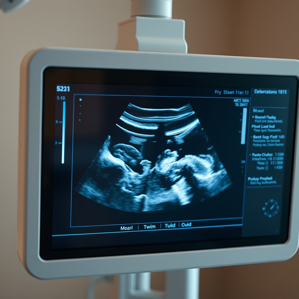
Ultrasound is the primary tool for spotting TTTS and tracking its progression.
What is twin‑twin transfusion syndrome?
TTTS occurs when identical twins share a single placenta (monochorionic) and develop abnormal blood‑vessel connections—called vascular anastomoses—that shunt blood preferentially from one twin (the donor) to the other (the recipient). This imbalance leads to:
Recipient twin: excess blood volume, polyhydramnios (high amniotic fluid), and risk of heart overload.
The condition typically appears after 16 weeks, when the circulatory systems of the twins become more interdependent. The exact cause of the abnormal connections is not fully understood, but researchers believe early placental development and genetic factors play a role (ACOG Practice Bulletin 2020; NICE Guideline NG31, 2022).
Because the twins share circulation, any intervention that alters blood flow can affect both babies. That is why management decisions are made by a multidisciplinary team—maternal‑fetal medicine specialists, neonatologists, and pediatric cardiologists—all weighing the risks and benefits for each fetus.
Most families discover TTTS during a routine anatomy scan. The key to better outcomes is recognizing the pattern of fluid imbalance early, then moving quickly to a specialist center that offers fetoscopic laser therapy. Even when the diagnosis feels overwhelming, knowing the underlying mechanism helps demystify the next steps.
How the five TTTS stages are defined
Doctors use the Quintero staging system (named after Dr. Juan Quintero) to describe the severity of TTTS based on ultrasound findings. The stages guide both prognosis and treatment choice. Below is a concise overview of each stage, followed by a detailed table you can reference during appointments.
Stage I – Mild imbalance
Donor twin has oligohydramnios (maximum vertical pocket < 2 cm) and the recipient twin has polyhydramnios (maximum vertical pocket > 8 cm). Both twins have normal bladder filling and Doppler studies are reassuring.
Stage II – Worsening fluid imbalance
Same fluid findings as stage I, but the donor twin’s urinary bladder is no longer visible on ultrasound, indicating reduced renal perfusion.
Stage III – Doppler abnormalities
In addition to stage II findings, Doppler studies show abnormal blood flow: either reversed flow in the ductus venosus of the donor or pulsatile flow in the umbilical artery of the recipient.
Stage IV – Fetal hydrops
One or both twins develop hydrops (fluid accumulation in two or more compartments such as skin, abdomen, or pleura), signaling severe cardiac strain.
Stage V – End‑stage TTTS
One twin has died in utero (often the donor) while the other continues to grow. The surviving twin is at high risk for neurological injury.
Stage
Key Ultrasound Findings
Typical Gestational Age Range
Prognosis (without treatment)
I
Oligohydramnios <2 cm (donor) + polyhydramnios >8 cm (recipient); both bladders visible; normal Dopplers
16‑22 weeks
≈ 70 % survival of both twins
II
Donor bladder not seen; fluid imbalance persists; Dopplers normal
18‑24 weeks
≈ 45‑55 % survival of both twins
III
Abnormal ductus venosus or umbilical artery Dopplers; fluid imbalance
20‑26 weeks
≈ 30‑40 % survival of both twins
IV
Fetal hydrops in one or both twins
22‑28 weeks
≈ 15‑25 % survival of both twins
V
Intra‑uterine death of one twin
Any GA after 16 weeks
Survival of remaining twin varies; high risk of neuro‑developmental issues
When you first hear a stage number, it can feel like a label that defines your entire pregnancy. Remember, the stage is a snapshot—not a destiny. Intervention timing, especially relative to GA, can dramatically shift outcomes.
Because the stages are based on measurable ultrasound criteria, many families find it reassuring to track the numbers themselves. A clear picture of the fluid pockets, bladder visibility, and Doppler waveforms can turn an abstract diagnosis into concrete data you can discuss with your doctor.
Use the TTTS Quintero Staging calculator to see how your ultrasound numbers translate into a stage.
How gestational age guides treatment decisions
Gestational age (GA) is the single most influential factor when choosing a TTTS treatment. The developing placenta, fetal organs, and amniotic fluid dynamics all change week by week, affecting both the safety of procedures and the likelihood of success.
Before 18 weeks: The placenta is still maturing, and laser surgery carries higher risk of damaging vital vessels. Expectant management or amnioreduction is often preferred, with close monitoring.
18‑26 weeks (the “golden window”): This is when fetoscopic laser photocoagulation (FLP) shows the best outcomes—survival rates approach 60‑70 % for both twins (ACOG 2020). The procedure aims to seal the abnormal anastomoses while preserving enough placental tissue for growth.
After 26 weeks: The fetus is nearing term, so the focus may shift to stabilizing the pregnancy and planning early delivery, especially if hydrops or severe Doppler changes appear.
In addition to the week count, clinicians consider the crown‑rump length and the size of the amniotic fluid pockets. For example, a donor twin with severe oligohydramnios at 22 weeks may benefit from amnioreduction to relieve compression, while a 24‑week recipient with rising polyhydramnios and abnormal Dopplers would be a strong candidate for laser.
Because each case is unique, your care team will integrate GA, stage, and the twin’s individual health markers into a personalized plan. The goal is to intervene early enough to correct the blood‑flow imbalance, but not so early that the procedure itself creates new complications.
When you discuss GA with your provider, ask for the specific “window of opportunity” for laser in your situation, and whether any alternative timing (e.g., delayed laser with steroids for lung maturity) might be safer based on your twins’ growth curves.
Management options by stage
Below is a concise guide to the typical interventions for each Quintero stage, along with the role GA plays in selecting the most appropriate approach.
Stage I – Expectant management (often 16‑20 weeks)
Expectant monitoring: Weekly ultrasounds to track fluid levels and fetal growth. Many cases resolve spontaneously, especially when GA is <18 weeks.
Amnioreduction: If polyhydramnios causes maternal discomfort or cervical shortening, a small amount of fluid can be removed from the recipient sac.
When to consider laser: If fluid imbalance worsens or Doppler changes appear before 22 weeks, laser may be offered even at stage I.
In practice, expectant management means setting a clear schedule for scans and having a rapid‑response plan if any red‑flag signs appear. Parents often find comfort in a written “ultrasound calendar” that outlines when each scan will happen and what specific measurements will be reviewed.
Stage II – Amnioreduction vs. laser (usually 18‑24 weeks)
Amnioreduction: Repeated drainage (often 2‑3 sessions) can relieve pressure, improve donor bladder filling, and buy time for a later laser if needed.
Laser photocoagulation (FLP): Preferred when donor bladder remains absent and fluid imbalance persists. Success rates climb to ~60 % for both twins when performed between 18‑26 weeks.
Septostomy: Rarely used; involves creating a small opening between the amniotic sacs to equalize fluid. Considered only when laser is unavailable and amnioreduction fails.
Choosing between amnioreduction and laser often hinges on how quickly the fluid imbalance is progressing. If the donor’s bladder disappears for more than a week, many centers move straight to laser to prevent irreversible renal damage.
Stage III – Laser is standard (20‑26 weeks)
Fetoscopic laser photocoagulation: The definitive therapy. The surgeon inserts a tiny fiber‑optic scope through the mother’s abdomen, identifies the shared vessels, and coagulates them with a laser. The procedure typically lasts 60‑90 minutes.
Adjunctive amnioreduction: May be done immediately after laser if polyhydramnios remains severe.
Delivery planning: If laser is successful and both twins stabilize, the pregnancy is usually allowed to continue to at least 34 weeks, unless maternal complications develop.
After laser, the team will monitor for “recanalization”—the rare re‑formation of abnormal vessels. This is why a follow‑up scan at 7‑10 days is standard, to confirm that the laser spots have sealed the connections.
Stage IV – Aggressive intervention (22‑28 weeks)
Laser plus intensive monitoring: Even with hydrops, laser can reverse fluid overload in the recipient and improve donor output, though survival drops to ~25‑35 % for both twins.
Early delivery: If hydrops does not resolve within 1‑2 weeks post‑laser or if maternal health is threatened, delivery at 28‑30 weeks is considered, often followed by neonatal intensive care.
Supportive care: In cases where laser is not feasible, amnioreduction and maternal steroids for lung maturity are employed while preparing for preterm birth.
When hydrops is present, the neonatology team is usually consulted before the laser procedure so that they can be on standby for immediate post‑delivery care. This coordinated approach improves the odds that both babies receive the specialized support they need right after birth.
Stage V – Intra‑uterine death of one twin (any GA after 16 weeks)
Observation of surviving twin: The primary concern is preventing brain injury from sudden blood‑pressure shifts. Continuous Doppler surveillance is essential.
Laser for remaining twin: If the surviving twin is still sharing vessels, laser may be performed to protect it from further hemodynamic stress.
Delivery timing: Many clinicians aim for delivery at 34‑36 weeks to minimize prematurity while allowing maximal fetal growth.
Even after the loss of one twin, the surviving fetus can thrive with careful monitoring. Parents are encouraged to discuss neuro‑protective strategies, such as magnesium sulfate administration, which is recommended by ACOG for neuroprotection in preterm deliveries.
Each intervention carries its own set of risks—bleeding, infection, preterm labor, or inadvertent damage to healthy placental vessels. Your team will discuss these in detail, weighing them against the potential benefit of preserving both lives.
Monitoring protocols before and after intervention
Even the most skilled surgeon cannot guarantee a perfect outcome without vigilant follow‑up. Monitoring schedules differ by stage and treatment, but the core elements are consistent:
Ultrasound frequency:
Stage I–II expectant: every 1‑2 weeks.
Post‑laser (any stage): every 3‑5 days for the first two weeks, then weekly.
Doppler studies: Umbilical artery, ductus venosus, and middle cerebral artery velocities are checked at each scan to catch early signs of cardiac strain.
Maternal symptoms: New or worsening abdominal pain, rapid weight gain, or sudden shortness of breath should prompt immediate evaluation.
Blood tests: Maternal hemoglobin and inflammatory markers are monitored after invasive procedures to rule out infection.
Fetal heart monitoring: Non‑stress tests (NST) or biophysical profiles (BPP) are added after 28 weeks to assess fetal well‑being.
After laser, the most critical early sign of success is the re‑appearance of the donor twin’s bladder and a normalization of amniotic fluid volumes. If fluid imbalance persists beyond 10 days, additional amnioreduction or a repeat laser may be considered.
Many centers now employ a “tele‑ultrasound” check‑in, where a sonographer transmits images to the fetal medicine team in real time. This can reduce travel burden for families living far from a tertiary center while still ensuring timely detection of any change.
Outcomes and survival rates by stage and gestational age
Survival statistics are averages; individual outcomes can vary widely based on the precise anatomy of the shared vessels, the skill of the surgical team, and the timing of intervention.
Stage
GA at Treatment
Combined Twin Survival (no laser)
Combined Twin Survival (laser)
Donor‑Twin Survival (laser)
Recipient‑Twin Survival (laser)
I
16‑20 weeks
≈ 70 %
≈ 80 %
≈ 85 %
≈ 85 %
II
18‑24 weeks
≈ 45‑55 %
≈ 60‑70 %
≈ 70 %
≈ 70 %
III
20‑26 weeks
≈ 30‑40 %
≈ 55‑65 %
≈ 65 %
≈ 65 %
IV
22‑28 weeks
≈ 15‑25 %
≈ 30‑40 %
≈ 40 %
≈ 40 %
V
any
≈ 10‑20 % (both)
≈ 45‑55 % (surviving twin)
—
≈ 55 %
Key take‑aways from the data:
Laser therapy consistently improves combined survival by 15‑30 % across stages.
Survival gaps narrow as GA advances; after 30 weeks, the advantage of laser diminishes because the twins are nearing term.
Donor‑twin survival is historically lower, reflecting the greater vulnerability to low perfusion. Early intervention helps close that gap.
These numbers are drawn from multicenter studies endorsed by the Society for Maternal‑Fetal Medicine (SMFM) and the European Twin Registry (2021). They align with ACOG’s recommendations that laser should be offered to any TTTS case from stage II onward when GA is between 18‑26 weeks.
It’s also worth noting that survival statistics are improving as technology advances. Newer laser fibers with finer precision and real‑time 3‑D imaging have reduced operative times and complication rates in recent trials.
Postnatal care and follow‑up considerations
Even after a successful delivery, twins who have experienced TTTS need coordinated neonatal and long‑term care.
Neonatal intensive care: Preterm twins (often 28‑34 weeks) may require ventilation, surfactant therapy, and careful fluid management. The recipient twin is monitored for cardiac overload, while the donor twin is watched for growth restriction.
Neuro‑developmental screening: Both twins should have cranial ultrasound or MRI in the first weeks of life, followed by developmental assessments at 6, 12, and 24 months.
Growth tracking: Serial weight, length, and head‑circumference measurements are crucial, as donor twins may have lingering growth challenges.
Cardiology follow‑up: The recipient twin often needs echocardiograms to ensure that any prenatal cardiac strain has resolved.
Parental support: Counseling and support groups for families who have experienced TTTS can help address anxiety, grief, and bonding challenges.
Because TTTS can affect the placenta’s overall function, clinicians also monitor the mother’s postpartum recovery for signs of retained placental tissue or infection.
Long‑term, many children born after TTTS have normal neurodevelopment, especially when they receive early intervention services if any delays are noted. Families are encouraged to keep a “developmental diary” to track milestones and share it with pediatricians during well‑child visits.
From our medical team: TTTS is a complex, high‑stakes condition, but it is also one where timely, stage‑appropriate treatment can dramatically improve outcomes. If you’re facing a diagnosis, lean on your maternal‑fetal medicine specialist to explain the exact numbers, the GA‑specific options, and the realistic expectations for each twin. Ask for a written summary of the plan, and don’t hesitate to seek a second opinion if anything feels unclear.
Emotional and psychological support for families
A TTTS diagnosis can feel like an emotional earthquake. The uncertainty surrounding the twins’ health, the need for frequent hospital visits, and the possibility of early delivery create a high‑stress environment. Recognizing the emotional toll is the first step toward coping.
Many hospitals now embed a perinatal mental‑health professional within the fetal‑medicine team. These specialists can provide brief counseling, teach grounding techniques, and help families process grief if one twin does not survive. Studies from the NHS (2022) show that families who receive structured emotional support report lower anxiety scores and better adherence to monitoring schedules.
Support groups—both in‑person and online—are invaluable. Hearing other parents describe similar night‑time ultrasound anxieties, the “waiting for the next scan” feeling, and the relief of seeing the donor’s bladder reappear can normalize your experience and reduce isolation.
Practical self‑care tips include: scheduling “quiet evenings” after each scan, keeping a journal of questions to ask your provider, and setting realistic expectations for sleep and nutrition. Remember, caring for your own wellbeing directly benefits the twins.
Placental pathology and implications for future pregnancies
TTTS is fundamentally a placental disorder. After delivery, the placenta is examined to understand the pattern of vascular anastomoses that caused the transfusion. Pathology reports often describe “arterio‑arterial” and “arterio‑venous” connections, as well as any areas of infarction or abnormal villous maturation.
For families planning future pregnancies, the presence of a monochorionic placenta in a subsequent pregnancy does not guarantee recurrence, but the risk is higher than in the general population. The Royal College of Obstetricians and Gynaecologists (RCOG) recommends that women with a prior TTTS receive early detailed ultrasound (around 12‑14 weeks) in any later pregnancy to assess chorionicity and initiate close monitoring if monochorionic.
Some emerging research suggests that low‑dose aspirin (81 mg) before 12 weeks may improve placental blood flow in high‑risk monochorionic pregnancies, though definitive evidence is still pending. Discuss any prophylactic strategies with your provider before trying to conceive again.
Screening and early detection strategies
Early detection of TTTS hinges on accurate chorionicity assessment and timely ultrasound surveillance. The first‑trimester scan (11‑13 weeks) should confirm whether the twins are monochorionic‑diamniotic; this distinction determines the follow‑up schedule.
For confirmed monochorionic twins, the standard of care (per ACOG and NICE) is a detailed anatomy scan at 16‑18 weeks, with a focused assessment of amniotic fluid volumes, bladder visibility, and Doppler flow. Some centers add a “mid‑trimester check” at 20 weeks to catch early TTTS changes before they become severe.
In addition to routine ultrasounds, maternal serum markers are not reliable for TTTS detection, but emerging fetal‑MRI techniques may someday provide a non‑invasive view of placental vascular architecture. Until then, the most actionable step is to ensure you have a qualified fetal‑medicine sonographer performing each scan.
Ask your provider for a written “ultrasound timeline” that outlines when each scan will occur, what specific measurements will be taken, and what thresholds would trigger an urgent review. Having this roadmap can reduce anxiety and give you clear checkpoints to discuss with your care team.
Myth vs. fact
Myth: “If the ultrasound shows TTTS, both babies are doomed.”
Fact: Early detection and treatment, especially laser photocoagulation between 18‑26 weeks, can lead to survival of both twins in 60‑70 % of cases (ACOG 2020).
Myth: “Only laser surgery works; amnioreduction is useless.”
Fact: Amnioreduction can relieve symptoms, buy time for a later laser, and is the preferred initial step in very early gestation (before 18 weeks) or when laser is not immediately available.
Myth: “TTTS always progresses to stage V.”
Fact: Many pregnancies stabilize after treatment, and some stage I cases resolve spontaneously without progressing.
Key takeaways
TTTS is a placental blood‑flow disorder that progresses through five Quintero stages, each with specific ultrasound criteria.
Gestational age (18‑26 weeks) is the critical window for laser photocoagulation, the most effective treatment for stages II‑III.
Stage I may be managed expectantly; stage II often benefits from amnioreduction or early laser; stage III‑IV usually require laser plus close monitoring; stage V focuses on protecting the surviving twin.
Survival rates improve by roughly 15‑30 % with laser across all stages, but outcomes still depend on exact GA and twin health.
Post‑delivery care includes NICU support, cardiac follow‑up, and neuro‑developmental screening for both twins.
Always keep a clear line of communication with your care team—ask for written summaries, use the TTTS Quintero Staging calculator, and report any new symptoms promptly.
Frequently asked questions
What is twin‑twin transfusion syndrome?
TTTS is a condition in monochorionic‑diamniotic twins where abnormal blood‑vessel connections cause one twin (the donor) to lose fluid and blood, while the other (the recipient) gains excess fluid and blood, leading to imbalanced growth and potential organ damage.
How are the stages of TTTS determined?
The stages are defined by the Quintero system, which looks at amniotic fluid volumes, visibility of each twin’s bladder, and Doppler blood‑flow patterns; each stage reflects increasing severity from mild fluid imbalance (stage I) to fetal hydrops (stage IV) and intra‑uterine death of one twin (stage V).
When is laser surgery recommended for TTTS?
Laser photocoagulation is typically recommended for stage II or higher when gestational age is between 18 and 26 weeks, especially if Doppler abnormalities appear or fluid imbalance persists despite amnioreduction.
Can TTTS resolve on its own without treatment?
Some stage I cases may improve spontaneously, particularly before 18 weeks, but most TTTS cases progress without intervention; close monitoring is essential to catch any worsening early.
What are the risks of TTTS for the donor twin?
The donor twin is at higher risk for growth restriction, oligohydramnios, and renal impairment because of reduced blood flow; untreated, donor‑twin survival can drop below 30 % in later stages.
How does gestational age affect TTTS treatment decisions?
Gestational age determines the safety and efficacy of interventions: early (<18 weeks) favors expectant or amnioreduction management; 18‑26 weeks is the optimal window for laser; after 26 weeks, the focus may shift to stabilizing the twins and planning early delivery.
What follow‑up care is needed for the surviving twin after stage V?
After the loss of one twin, the surviving twin is monitored with frequent Doppler studies, and many teams give a course of magnesium sulfate for neuro‑protection. Delivery is usually planned at 34‑36 weeks to balance prematurity risks with continued growth.
Can lifestyle changes, like nutrition or activity, influence TTTS outcomes?
While no specific diet can prevent TTTS, maintaining a balanced prenatal diet and staying hydrated support overall placental health. Your provider may recommend moderate activity and avoiding heavy lifting, especially after invasive procedures like laser.
When to call your doctor
If you notice any of the following, contact your obstetrician or midwife immediately: sudden increase in abdominal girth, severe abdominal pain, vaginal bleeding, rapid weight gain, decreased fetal movements, or signs of preterm labor (regular contractions, cervical change). This article is for informational purposes only and does not replace personalized medical advice.
References
American College of Obstetricians and Gynecologists (ACOG). Practice Bulletin No. 197: Twin‑Twin Transfusion Syndrome. 2020.
National Institute for Health and Care Excellence (NICE). Twin‑Twin Transfusion Syndrome: Diagnosis and Management (NG31). 2022.
Society for Maternal‑Fetal Medicine (SMFM). Clinical Management Guidelines for Monochorionic Twins. 2021.
European Twin Registry. Outcomes of Fetoscopic Laser Photocoagulation for TTTS. 2021.
Centers for Disease Control and Prevention (CDC). Congenital Anomalies and Twin Pregnancies. 2023.
World Health Organization (WHO). Maternal and Neonatal Health: Placental Disorders. 2022.
When Shubhra Mishra was expecting her first child in 2016, she was overwhelmed by conflicting food advice — one site said yes, another said never. By the time her second baby arrived in 2019, she realized millions of mothers face the same confusion.
That sparked a five-year journey through clinical nutrition papers, cultural diets, and expert conversations — all leading to BumpBites: a calm, compassionate space where science meets everyday motherhood.
Her long-term vision is to build a global community ensuring safe, supported, and free deliveriesfor every mother — because no woman should face pregnancy alone or uninformed. 🌿
🌍 Stand with mothers, shape safer guidance
Join a small circle of experts who review BumpBites articles so expecting parents everywhere can decide with confidence.