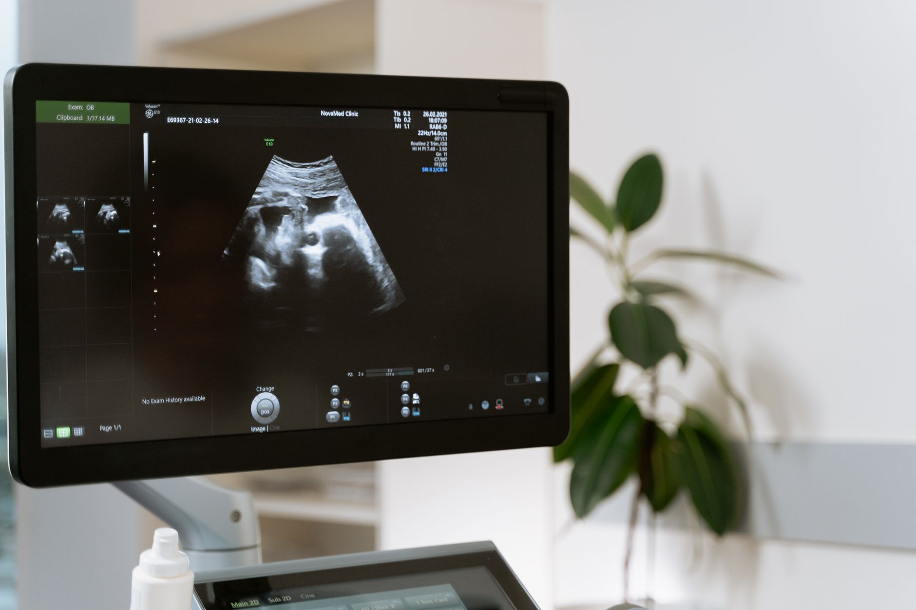
A third trimester pregnancy ultrasound checks baby’s growth, position, and health. Learn what to expect, why it’s done, and how to prepare for this important scan.
By Shubhra Mishra — a mom of two who turned her own confusion during pregnancy into BumpBites, a global mission to make food choices clear, safe, and stress-free for every expecting mother. 💛
Check whether any food is safe during pregnancy with the BumpBites Food Safety Checker.
Quick take: A third‑trimester pregnancy ultrasound is safe for both baby and mother, helps monitor growth, placenta position, amniotic fluid, and fetal presentation, and is typically scheduled between 28 and 36 weeks. It usually lasts 15–30 minutes, costs around $200‑$400 without insurance in the U.S., and requires no special preparation beyond a full bladder.
It’s 10 p.m., you’re curled up on the couch, and a sudden kick makes you wonder: “Do I need another scan before the due date?” You’re not alone—many expectant parents reach for their phones at the same moment, hoping for a clear answer. The short answer is yes, a third‑trimester ultrasound can provide valuable information, and it’s generally considered safe. In the sections below we’ll walk through what you’ll actually see, how the scan works, when to schedule it, what the numbers mean, and how to prepare so the experience feels as calm as possible.
We’ll also cover practical details like cost, typical duration, and what to ask your provider. By the end of this guide you’ll have a checklist in hand, know the key differences between 2D and 3D imaging, and feel confident discussing any findings with your care team.
Seeing the baby’s movements on the screen can turn anxiety into reassurance.
What can you see in a third trimester pregnancy ultrasound?
By the third trimester the baby’s major organs are already formed, so the scan focuses on growth and positioning rather than structural anomalies. Typical views include:
Placenta location—whether it’s low‑lying, covering the cervical opening (placenta previa), or attached normally.
Amniotic fluid volume—assessed with the deepest vertical pocket (DVP) or the amniotic fluid index (AFI).
Fetal presentation—head‑down (cephalic), breech, or transverse.
Doppler flow—blood‑flow patterns in the umbilical artery and middle cerebral artery, useful for assessing placental efficiency.
Because the baby’s bones are ossified, you’ll also see clearer skeletal outlines, which can help confirm the estimated weight and detect any late‑onset growth restriction.
How safe is a third trimester ultrasound for baby and mother?
U
ltrasound uses sound waves, not ionizing radiation, and the American College of Obstetricians and Gynecologists (ACOG) states that “diagnostic ultrasound performed by a qualified provider is safe when used according to established guidelines.” The thermal and mechanical indices are kept below levels that could cause tissue heating. Studies reviewed by the World Health Organization (WHO) have found no link between routine third‑trimester scans and adverse fetal outcomes.
That said, the FDA recommends keeping the ALARA principle—“as low as reasonably achievable”—in mind. Technicians limit the scan time and avoid prolonged exposure to a single area. For the mother, there is no known increase in miscarriage risk, and no contraindications for typical pregnancies. Women with certain rare conditions (e.g., severe abdominal scarring) may be advised to use alternative imaging, but this is the exception rather than the rule.
When should you schedule a third trimester ultrasound appointment?
Most providers recommend a growth scan between 28 and 32 weeks, and a final assessment around 36 weeks if there are concerns about fetal size, placenta position, or amniotic fluid. ACOG’s practice bulletin suggests:
28‑32 weeks—baseline growth assessment and placental location.
34‑36 weeks—follow‑up if earlier scans showed borderline measurements or if the mother has risk factors such as hypertension or diabetes.
38‑40 weeks—optional scan to confirm presentation and fluid when labor is imminent.
If you’re in a high‑risk pregnancy (e.g., multiple gestation, pre‑eclampsia), your clinician may schedule more frequent scans. Always check with your provider whether a referral is needed; in many U.S. states a physician order is required for insurance coverage, but private clinics often accept self‑referral.
What signs of placenta previa appear on a third trimester ultrasound?
Placenta previa is identified when the placenta partially or completely covers the internal cervical os. On the screen you’ll see a bright, rounded structure hugging the lower uterine segment. Key sonographic signs include:
Low‑lying placenta—within 2 cm of the cervical os.
Complete coverage—the placenta entirely overlies the os.
Marginal previa—edge of the placenta touches but does not cover the os.
If a low‑lying placenta is detected, ACOG recommends repeat ultrasound at 28‑32 weeks because many placentas “migrate” upward as the uterus expands. Persistent previa after 36 weeks often leads to a planned cesarean delivery to avoid painless bleeding.
How to interpret fetal growth measurements in a third trimester ultrasound?
Growth is assessed by comparing measured biometry to standardized percentile charts (e.g., Hadlock formulas). The key steps are:
Measure HC, AC, FL, and calculate EFW.
Plot each measurement on gestational‑age‑specific curves.
Identify if the EFW falls below the 10th percentile (potential growth restriction) or above the 90th percentile (possible macrosomia).
For example, an AC of 33 cm at 32 weeks corresponds to roughly the 50th percentile, indicating average growth. If the AC were 28 cm, that would place the baby near the 10th percentile, prompting closer monitoring of Doppler flow and maternal health.
Clinicians also look at the growth velocity—how much the measurements have changed since the last scan. A slowdown may signal placental insufficiency even if the absolute size is still within normal limits.
Differences between 2D and 3D ultrasounds in the third trimester
Both modalities are safe, but they serve different purposes. 2D ultrasound provides real‑time grayscale images, ideal for measurements and Doppler flow. 3D adds a volumetric reconstruction that can be rotated, offering clearer views of surface anatomy—useful for parental bonding and sometimes for evaluating facial anomalies.
Feature
2D Ultrasound
3D Ultrasound
Doppler (often combined with 2D)
Primary use
Biometry, growth, placenta, fluid
Surface anatomy, parental keepsakes
Blood‑flow assessment
Image type
Flat, real‑time slices
Volumetric, rotatable images
Color/Power flow maps
Scan time
10‑20 min
15‑30 min (includes processing)
5‑10 min added to 2D
Cost impact
Standard fee
+$100‑$200 extra
Usually included in standard fee
Clinical necessity
Essential for all third‑trimester checks
Optional, mainly for maternal‑fetal bonding
Critical for high‑risk pregnancies
In most routine third‑trimester assessments, 2D plus Doppler provides all the data needed. 3D is a nice add‑on if you want a keepsake or if a facial anomaly is suspected, but it does not replace the standard measurements.
Can a third trimester ultrasound detect birth defects?
By the third trimester many structural anomalies have already been identified on the anatomy scan (typically performed at 18‑22 weeks). However, a third‑trimester scan can still spot late‑appearing issues such as:
Severe hydrocephalus (enlarged ventricles).
Significant limb abnormalities that become more apparent as the fetus grows.
Major facial clefts that were subtle earlier.
Because the bones are ossified, the resolution for subtle cardiac defects diminishes compared with the earlier anatomy scan. If a defect is suspected, your provider may recommend a detailed fetal echocardiogram or MRI instead of a routine third‑trimester scan.
How long does a third trimester ultrasound typically last?
Most third‑trimester scans last between 15 and 30 minutes. The exact duration depends on:
Whether 3D imaging is requested (adds ~5‑10 minutes).
Complexity of the case—e.g., if Doppler studies are needed.
Maternal factors such as body habitus, which can affect image acquisition.
Technicians aim to keep the total exposure time well under the safety thresholds set by the FDA and ACOG, so you’ll rarely be in the room for more than half an hour.
Third trimester ultrasound cost without insurance
In the United States, out‑of‑pocket fees range from $200 to $400 for a standard 2D scan. Adding 3D imaging can increase the price by $100‑$200. In Canada and the United Kingdom, publicly funded health systems (e.g., NHS) cover the scan at no cost for patients who meet clinical criteria. Private clinics may charge similar fees to the U.S., but many offer package discounts if you bundle the scan with other prenatal services.
When budgeting, remember that additional Doppler studies or repeat scans for growth monitoring may be billed separately. Always ask the clinic for a detailed estimate before you schedule the appointment.
What to expect during a third trimester ultrasound exam
When you arrive, you’ll be asked to fill out a brief health questionnaire. You’ll then be taken to a dimly lit room with a reclining chair. The technician will place a warm, water‑based gel on your abdomen and use a transducer to capture images. You’ll be instructed to drink about 32 oz (1 liter) of water 30‑45 minutes before the scan to ensure a full bladder, which provides a clear acoustic window.
During the exam you’ll hear a soft hum as the probe moves across your belly. The technician may ask you to hold your breath briefly to improve image quality. You’ll be able to watch the monitor in real time, and many clinics invite you to view the images on a tablet or screen. After the scan, a written report is generated and sent to your obstetrician.
Third trimester ultrasound checklist for expectant mothers
Having a short list can make the appointment feel smoother. Here’s a practical checklist:
Drink 2‑3 glasses of water 30 minutes before the visit.
Wear comfortable, loose‑fitting clothing (e.g., a maternity top that can be lifted).
Bring your insurance card and a photo ID.
Prepare a list of questions (e.g., “Is my baby’s growth on track?”).
Bring a notebook or phone to record the provider’s explanations.
Ask about the possibility of a 3D image if you’d like a keepsake.
How often should a third trimester ultrasound be done?
For low‑risk pregnancies, a single growth scan at 28‑32 weeks is usually sufficient. High‑risk cases—such as maternal hypertension, diabetes, or a history of intrauterine growth restriction—may require serial scans every 2‑4 weeks. ACOG’s guidelines suggest:
Low risk—one scan unless clinical concerns arise.
Moderate risk—additional scans at 34 weeks and 38 weeks.
High risk—every 2‑3 weeks from 28 weeks onward.
Always follow your provider’s individualized plan; they’ll tailor the frequency based on your health and any prior findings.
Third trimester ultrasound and amniotic fluid levels
Amniotic fluid volume is a key marker of fetal well‑being. The sonographer measures the deepest vertical pocket (DVP); a value <2 cm may indicate oligohydramnios (low fluid), while >8 cm suggests polyhydramnios (excess fluid). Both conditions can affect labor and may signal underlying issues such as placental insufficiency or maternal diabetes.
If abnormal fluid levels are detected, your provider may recommend:
More frequent monitoring (every 1‑2 weeks).
Maternal hydration strategies.
Early delivery if the situation threatens the baby’s health.
These decisions are guided by ACOG’s recommendations and tailored to each pregnancy.
Third trimester ultrasound and fetal position for delivery
At 36‑38 weeks the sonographer will note the baby’s presentation: cephalic (head‑down), breech, or transverse. A breech presentation occurs in about 3‑4 % of term pregnancies. If breech is identified, your provider may suggest a version‑attempt (external cephalic version) or plan for a cesarean delivery, especially if the baby is not turning on its own.
Even when the baby is head‑down, the exact angle (e.g., left occiput anterior) can influence labor progress. Some providers use this information to anticipate potential shoulder dystocia or to decide on a trial of labor after cesarean (TOLAC).
Third trimester ultrasound vs. anatomy scan differences
The anatomy scan, performed at 18‑22 weeks, is a comprehensive structural survey focused on organ development. By contrast, the third‑trimester scan emphasizes:
Growth assessment—biometry to estimate weight.
Placental location—detecting previa or accreta.
Amniotic fluid—ensuring adequate volume.
Fetal presentation—preparing for delivery.
Both scans use the same equipment, but the goals shift from “are the organs formed correctly?” to “is the baby thriving and ready for birth?”
Biometry measurements help estimate the baby’s weight and growth trajectory.
From our medical team: Third‑trimester ultrasounds are a routine safety check, not a diagnostic test for every condition. If your scan shows something unexpected, we’ll discuss next steps and reassure you that most findings are manageable with close monitoring. Always bring any concerns to your next prenatal visit, and remember that each scan adds a piece to the larger picture of your pregnancy’s health.
Myth vs. fact
Myth: A third‑trimester ultrasound can harm the baby because it uses “radiation.”
Fact: Ultrasound uses sound waves, not ionizing radiation, and reputable guidelines confirm it is safe when performed by qualified professionals.
Myth: If the third‑trimester scan looks normal, there’s no need for any further monitoring.
Fact: Even with a normal scan, high‑risk conditions (e.g., gestational diabetes) may still require additional testing or more frequent visits.
Myth: You must have a full bladder for the entire scan, which is uncomfortable and risky.
Fact: A full bladder improves image quality, but technicians will pause if you feel uncomfortable, and the bladder is emptied after the exam.
Key takeaways
Third‑trimester ultrasounds are safe, non‑invasive, and primarily assess growth, placenta, fluid, and presentation.
Schedule the first growth scan between 28‑32 weeks; additional scans depend on risk factors.
Typical cost without insurance: $200‑$400 for 2D; add $100‑$200 for 3D if desired.
Expect the exam to last 15‑30 minutes; a full bladder helps obtain clearer images.
Bring a water bottle, comfortable clothing, and a list of questions to make the visit smooth.
Contact your provider promptly if you notice decreased fetal movement, severe abdominal pain, or bleeding.
Frequently asked questions
Is a third trimester ultrasound safe for the baby?
Yes. Ultrasound uses sound waves, not radiation, and professional guidelines (ACOG, WHO) confirm it is safe when performed according to standard protocols.
What can a third trimester ultrasound show that earlier scans cannot?
It provides real‑time data on fetal growth, amniotic fluid volume, placenta location, and presentation—information crucial for planning delivery that earlier anatomy scans don’t capture.
How long does a third trimester ultrasound take?
Typically 15‑30 minutes, depending on whether 3D imaging or Doppler studies are added.
Do I need a doctor’s referral for a third trimester ultrasound?
In most U.S. states, insurance requires a physician order, but many private clinics accept self‑referral. In the UK and Canada, the scan is usually arranged through your obstetrician or midwife.
Can a third trimester ultrasound predict the baby’s birth weight?
It estimates fetal weight using biometry formulas, which are accurate within ±10‑15 % for most babies. The prediction helps anticipate delivery method but isn’t a guarantee.
What are the signs of complications that can be seen on a third trimester ultrasound?
Findings such as low amniotic fluid, placenta previa, abnormal Doppler flow, or growth restriction signal potential complications and prompt closer monitoring or early delivery planning.
When to call your doctor
If you notice any of the following, contact your provider or go to the nearest emergency department immediately: sudden or severe abdominal pain, vaginal bleeding, a noticeable decrease in fetal movement, fever over 100.4°F (38°C), or fluid leaking from the vagina. This article is for informational purposes only and does not replace personalized medical advice.
References
American College of Obstetricians and Gynecologists. “Ultrasound Guidance for Obstetric Care.” ACOG Practice Bulletin No. 199, 2021.
World Health Organization. “Recommendations on Antenatal Care for a Positive Pregnancy Experience.” WHO Guidelines, 2022.
National Institute for Health and Care Excellence (NICE). “Routine Antenatal Care.” NICE Clinical Guideline CG62, 2023.
U.S. Food and Drug Administration. “Ultrasound Safety.” FDA Consumer Health Information, 2022.
Royal College of Obstetricians and Gynaecologists (RCOG). “Placenta Previa.” Green‑top Guideline No. 27, 2020.
Society for Maternal-Fetal Medicine. “Management of Suspected Fetal Growth Restriction.” SMFM Consensus Statement, 2021.
Mayo Clinic. “Third Trimester Ultrasound: What to Expect.” Patient Care Articles, 2023.
Centers for Disease Control and Prevention. “Prenatal Care.” CDC Health Information, 2022.
Not sure about the label on Third Trimester Pregnancy Ultrasound products?
Snap the ingredients list and SafeFilter checks every ingredient for your stage — only 3 free scans this month, then you're locked until reset. Unlimited from $7/mo or $50/yr.
When Shubhra Mishra was expecting her first child in 2016, she was overwhelmed by conflicting food advice — one site said yes, another said never. By the time her second baby arrived in 2019, she realized millions of mothers face the same confusion.
That sparked a five-year journey through clinical nutrition papers, cultural diets, and expert conversations — all leading to BumpBites: a calm, compassionate space where science meets everyday motherhood.
Her long-term vision is to build a global community ensuring safe, supported, and free deliveriesfor every mother — because no woman should face pregnancy alone or uninformed. 🌿
🌍 Stand with mothers, shape safer guidance
Join a small circle of experts who review BumpBites articles so expecting parents everywhere can decide with confidence.