

Quick take: Your first-trimester ultrasound—usually done between 6 and 12 weeks—is a safe, painless way to confirm your pregnancy, check your baby’s heartbeat, and estimate your due date. Most women see a tiny flicker of a heartbeat around 6–7 weeks, and the whole appointment takes about 15–30 minutes. It’s not the same as the nuchal translucency scan (that’s later, at 11–14 weeks), and you won’t find out the baby’s gender yet. Think of it as a first hello, not a full anatomy check.
You’re sitting in the dim ultrasound room, the gel still cool on your belly, heart pounding. The screen flickers, and suddenly—there it is. A tiny, rhythmic pulse. Your baby’s heartbeat. For many women, this moment is the first real proof that the little life inside is growing, safe, and on track.
But if you’re new to pregnancy, the idea of a first-trimester ultrasound can feel overwhelming. When should you schedule it? What exactly will the technician look for? Will it hurt? And most importantly—is it safe for your baby?
This guide covers everything you need to know about your first-trimester ultrasound: what to expect, how to prepare, what the scan measures, and why it matters. We’ll walk you through the procedure step by step, so you can walk into that appointment feeling calm, informed, and ready.
What to expect during a first trimester pregnancy ultrasound
You’ve probably seen ultrasound images—those grainy black-and-white photos of babies curled up in the womb. But what actually happens during the scan?
First, you’ll lie down on an exam table, and the technician (called a sonographer) will ask you to lift your shirt and lower your waistband slightly. They’ll squirt a clear, cool gel on your belly—this helps the ultrasound probe (called a transducer) glide smoothly and pick up clearer images. If you’re very early in pregnancy (around 6–8 weeks), they might use a transvaginal probe instead (a slender wand inserted into the vagina), which gives a closer look at the tiny embryo.
“I was so nervous about the transvaginal scan,” says Sarah, a first-time mom we spoke with. “But it was over in minutes, and honestly, it didn’t hurt at all. The technician was so gentle, and seeing that little heartbeat made it all worth it.”
The sonographer will move the probe around your belly, taking measurements and snapping images. You’ll see the screen, but it might look like a blur of gray and black at first. Don’t worry—they’ll point out what you’re seeing, like the gestational sac (the fluid-filled space where your baby is growing), the yolk sac (which nourishes the embryo early on), and, if you’re far enough along, the baby’s heartbeat.
The whole process is painless, though you might feel a little pressure from the probe. Some women feel emotional—seeing your baby for the first time can be overwhelming. Others feel relief, especially if they’ve had early pregnancy bleeding or cramping. “I burst into tears when I saw the heartbeat,” says Maria, another mom. “All the worry just melted away.”
How early can you see a fetal heartbeat in the first trimester?
With a transvaginal ultrasound (the more sensitive method used in early pregnancy), a heartbeat is usually visible by 6 weeks. At this stage, it looks like a tiny flicker on the screen—often too small to hear, but you can see it beating. By 7 weeks, the heartbeat is more distinct, and the sonographer can measure its rate (usually between 110 and 160 beats per minute).
With a transabdominal ultrasound (the probe on your belly), you’ll typically need to be at least 7–8 weeks to see a heartbeat. Before that, the embryo is too small for the probe to pick up clearly.
If you’re earlier than 6 weeks, you might only see the gestational sac—a small black circle on the screen. That’s normal! The embryo and heartbeat develop a little later. If your provider doesn’t see a heartbeat by 7 weeks, they might ask you to come back in a week or two to check again.
“I was 5 weeks and 5 days when I had my first ultrasound,” says Priya, a mom of two. “All I saw was the gestational sac, and I panicked. But my doctor reassured me it was too early for a heartbeat. When I went back at 7 weeks, there it was—beating strong.”

First trimester ultrasound vs. nuchal translucency scan: What’s the difference?
If you’ve heard terms like “dating scan” and “nuchal translucency scan,” you might be wondering: Aren’t all ultrasounds the same? Not quite. While both are done in the first trimester, they serve different purposes.
Here’s a quick breakdown:
| Feature | First Trimester Ultrasound (Dating Scan) | Nuchal Translucency Scan |
|---|---|---|
| When it’s done | 6–12 weeks | 11–14 weeks |
| Purpose | Confirm pregnancy, check heartbeat, estimate due date | Screen for chromosomal conditions (like Down syndrome) |
| What it measures | Gestational sac, embryo size, heartbeat | Nuchal translucency (fluid at the back of baby’s neck), nasal bone, blood flow |
| Type of ultrasound | Transvaginal or transabdominal | Transabdominal only |
| Do you need a full bladder? | Sometimes (if transabdominal) | Yes |
| Is it routine? | Yes, for most women | Optional (but recommended for high-risk pregnancies) |
The first-trimester ultrasound (often called the “dating scan”) is usually your first ultrasound. It confirms that the pregnancy is in the right place (not ectopic), checks for a heartbeat, and gives you a more accurate due date based on the baby’s size. It’s a quick, reassuring check-in.
The nuchal translucency scan, on the other hand, is a more detailed screening. It measures the fluid at the back of your baby’s neck—a marker that can indicate a higher risk of chromosomal conditions like Down syndrome or trisomy 18. This scan is usually combined with a blood test (called the combined screening) for more accuracy. It’s not diagnostic (meaning it doesn’t give a definite “yes” or “no”), but it can tell you if you’re at higher risk and might need further testing, like an amniocentesis or NIPT (non-invasive prenatal testing).
“I had both scans, and they felt very different,” says Aisha, a mom who opted for the nuchal translucency screening. “The first one was all about the excitement of seeing the baby. The second one felt more serious—like we were getting important information about our baby’s health.”
Is a first trimester ultrasound safe for the baby and mother?
This is the question that keeps many moms up at night: Is the ultrasound safe? The short answer: Yes, when done by a trained professional, first-trimester ultrasounds are considered very safe for both you and your baby.
Ultrasounds use high-frequency sound waves—not radiation, like X-rays—to create images of your baby. These sound waves bounce off structures in your body and are picked up by the probe, which then translates them into the images you see on the screen. There’s no evidence that this process harms a developing fetus.
The American College of Obstetricians and Gynecologists (ACOG), the National Health Service (NHS), and the World Health Organization (WHO) all agree that ultrasounds are safe when used appropriately. That means:
- They’re done by a trained sonographer or doctor.
- They’re done for a medical reason (not just for keepsake photos).
- They’re kept as short as possible (though your first-trimester scan is usually quick—just 10–20 minutes).
Some women worry about the heat generated by the ultrasound probe. While it’s true that prolonged exposure to high-intensity ultrasound can theoretically raise tissue temperature, the levels used in standard prenatal ultrasounds are well below the threshold considered risky. “The energy levels in diagnostic ultrasounds are very low,” explains Dr. Lisa Chen, an OB-GYN. “We’ve been using them for decades, and there’s no evidence they cause harm.”
That said, it’s always a good idea to avoid unnecessary ultrasounds—like those done for non-medical reasons (e.g., “keepsake” 3D/4D scans at commercial studios). Stick to the ultrasounds recommended by your provider, and you can feel confident that you’re doing what’s safest for your baby.
“I was so nervous about the ultrasound at first,” says Elena, a mom of three. “But my doctor explained that it’s been used safely for generations. Knowing that helped me relax and just enjoy seeing my baby for the first time.”
What does a first trimester ultrasound measure, and why?
You might be wondering: What exactly is the sonographer looking for during this scan? It’s not just about seeing the baby—it’s about gathering key information to make sure your pregnancy is off to a healthy start.
Here’s what your first-trimester ultrasound measures, and why each detail matters:
1. Gestational sac
The gestational sac is the first structure visible on an ultrasound—usually by 5 weeks. It’s the fluid-filled space where your baby will grow. Measuring the sac helps confirm that the pregnancy is in the right place (inside the uterus, not in the fallopian tubes, which would be an ectopic pregnancy).
2. Yolk sac
By 5–6 weeks, the yolk sac appears—a small circle inside the gestational sac. It provides nutrients to the embryo before the placenta takes over. Seeing the yolk sac is a good sign that the pregnancy is progressing normally.
3. Crown-rump length (CRL)
This is the most important measurement in the first trimester. The crown-rump length (CRL) is the distance from the top of your baby’s head to their bottom (like measuring a tiny curled-up shrimp). It’s used to:
- Estimate your due date (more accurate than using your last menstrual period, especially if your cycles are irregular).
- Check if the baby is growing at the expected rate.
- Screen for chromosomal conditions (if the CRL is smaller than expected, it might prompt further testing).
At 6 weeks, the CRL is usually about 2–4 mm. By 12 weeks, it’s around 5–6 cm.
4. Fetal heartbeat
As we mentioned earlier, the heartbeat is usually visible by 6–7 weeks. The sonographer will measure the heart rate (in beats per minute, or BPM). A normal first-trimester heartbeat is usually between 110 and 160 BPM. A very slow or very fast heartbeat can sometimes indicate a problem, but it’s not always a cause for concern—your provider will let you know if they want to monitor it further.
5. Number of babies
Yes, this is when you’ll find out if you’re having twins (or more!). The sonographer will check for multiple gestational sacs or embryos. If you are carrying multiples, they’ll note whether they share a placenta (monochorionic) or have separate ones (dichorionic), which affects how your pregnancy is monitored.
6. Uterus and ovaries
The ultrasound also checks your uterus and ovaries for any issues, like fibroids (non-cancerous growths) or ovarian cysts. These are common and usually harmless, but they might need to be monitored if they’re large or causing symptoms.
“I had no idea they were checking so many things,” says Jamie, a first-time mom. “When the technician pointed out the heartbeat and the crown-rump length, it made me realize how much is happening in those early weeks—even if I couldn’t feel it yet.”

How long does a first trimester pregnancy ultrasound appointment take?
You’re probably wondering: How much time should I block off for this? The good news is that first-trimester ultrasounds are usually quick—most appointments take 15 to 30 minutes, start to finish. Here’s a rough breakdown of what to expect:
- Check-in (5–10 minutes): You’ll fill out some paperwork (if it’s your first visit) and confirm your details with the receptionist.
- Prep (5 minutes): You might be asked to drink water and have a full bladder (more on that below). The sonographer will explain the process and answer any questions.
- The scan itself (10–20 minutes): This is the main event. The sonographer will take measurements, check the baby’s heartbeat, and snap some images. If you’re very early (6–7 weeks), they might use a transvaginal probe, which can take a little longer to get clear images.
- Wrap-up (5 minutes): The sonographer will print out some images for you to take home (your first baby photos!) and might give you a quick summary of what they saw. They won’t give you a full diagnosis—that’s up to your doctor or midwife, who will review the results and call you later.
If you’re having a nuchal translucency scan (the later first-trimester screening), the appointment might take a little longer—around 30–45 minutes. This is because it’s a more detailed scan, and the sonographer needs to take precise measurements of the fluid at the back of your baby’s neck.
“I was in and out in 20 minutes,” says Leah, a mom of one. “I thought it would take forever, but it was over before I knew it. The hardest part was waiting for my doctor to call with the results!”
Can you hear your baby’s heartbeat during a first trimester ultrasound?
This is one of the most magical moments of early pregnancy: hearing your baby’s heartbeat for the first time. But whether you’ll hear it during your first-trimester ultrasound depends on how far along you are—and what type of ultrasound you’re having.
Here’s what to expect:
Transvaginal ultrasound (6–8 weeks)
If you’re having a transvaginal ultrasound (the probe inserted into the vagina), you might not hear the heartbeat yet. At this stage, the heartbeat is often visible on the screen as a tiny flicker, but it’s usually too faint to pick up with the Doppler (the handheld device that amplifies the sound). Don’t worry—this doesn’t mean anything is wrong. It’s just too early for the sound to be strong enough.
Transabdominal ultrasound (8+ weeks)
By 8–10 weeks, if you’re having a transabdominal ultrasound (the probe on your belly), you might hear the heartbeat. The sonographer will turn on the Doppler, and you’ll hear a rapid, whooshing sound—like a tiny galloping horse. It’s unmistakable, and for many moms, it’s the moment when the pregnancy suddenly feels real.
“I’ll never forget the sound of my baby’s heartbeat at 9 weeks,” says Nina, a mom of two. “It was so fast—like a little drumroll. I burst into tears right there in the room.”
If you don’t hear the heartbeat during the ultrasound, don’t panic. It doesn’t necessarily mean there’s a problem. Sometimes the baby’s position makes it harder to pick up the sound, or the equipment isn’t sensitive enough. The sonographer will let you know if they’re concerned, and your provider will follow up with you after reviewing the images.
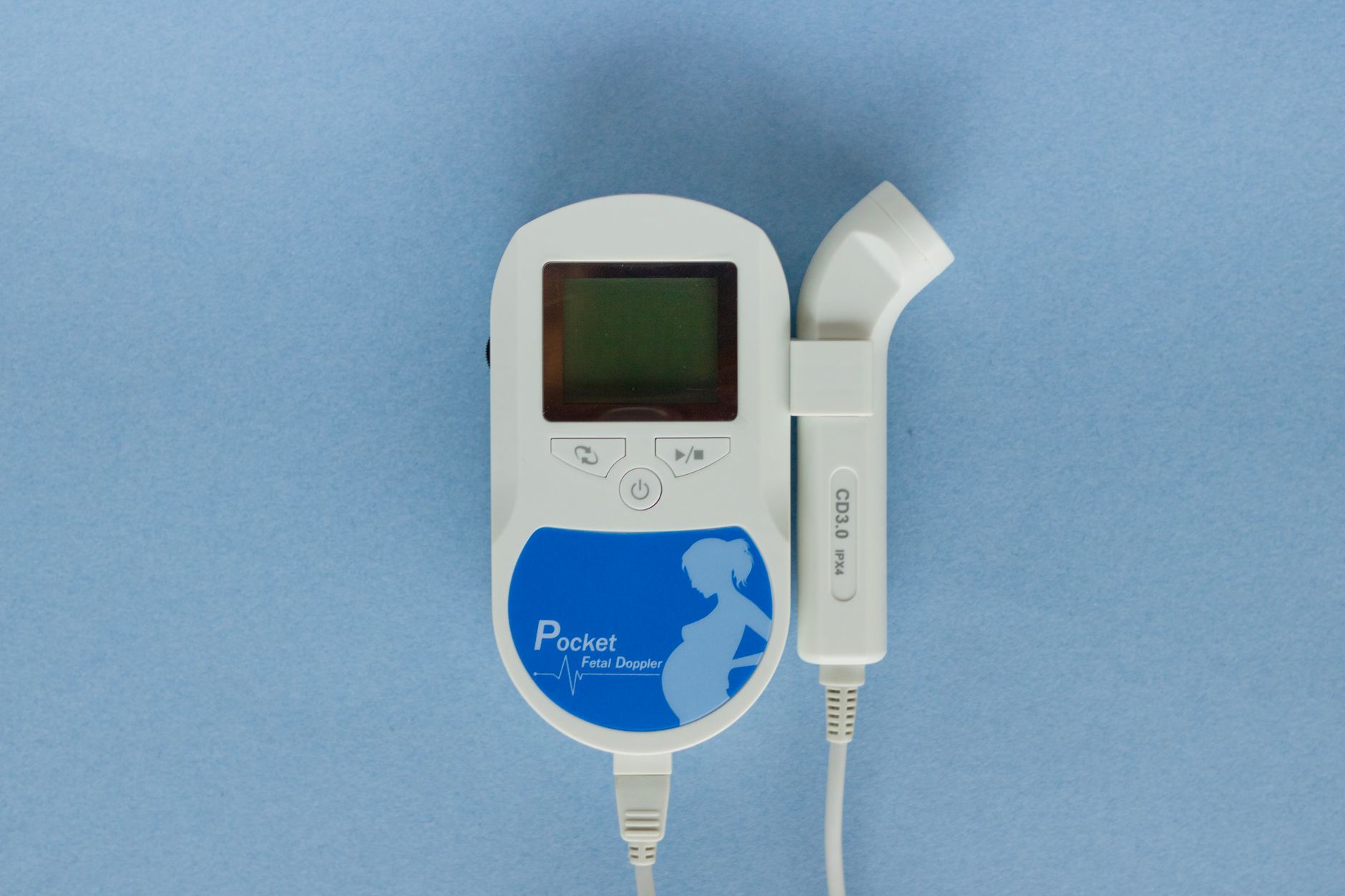
One thing to note: You won’t hear the heartbeat with a home Doppler this early. Those handheld devices (the ones you can buy online) aren’t sensitive enough to pick up a heartbeat until at least 12 weeks, and even then, it can be tricky. If you’re tempted to try one, talk to your provider first—they can cause unnecessary worry if you can’t find the heartbeat.
How much does a first trimester ultrasound cost without insurance?
Let’s talk about the practical side: How much will this cost? The price of a first-trimester ultrasound can vary a lot depending on where you live, where you go, and whether you have insurance. Here’s what you need to know:
With insurance
If you have health insurance, your first-trimester ultrasound is usually covered as a preventive service under the Affordable Care Act (in the U.S.). That means you might not pay anything out of pocket, or you might have a small copay (usually $20–$50). Check with your insurance provider to confirm your coverage—some plans require you to go to an in-network provider.
Without insurance
If you don’t have insurance, the cost can range from $100 to $500 or more. Here’s a rough breakdown of what you might pay:
- Obstetrician’s office or hospital: $200–$500 (this is usually the most expensive option).
- Independent ultrasound clinic: $100–$300 (often cheaper than hospitals, but make sure they’re reputable and send the results to your provider).
- Planned Parenthood or community health clinics: $50–$200 (sliding scale based on income).
- Mobile ultrasound services: $150–$400 (some companies come to your home, but be cautious—make sure they’re licensed and share results with your provider).
If cost is a concern, don’t hesitate to ask for a payment plan or discount. Many clinics offer financial assistance, especially if you’re paying out of pocket. You can also call around to compare prices—some places offer “self-pay” discounts if you pay upfront.
“I was uninsured when I had my first ultrasound,” says Megan, a mom who used a community clinic. “It was $120, but they let me pay in two installments. It was worth every penny to see my baby’s heartbeat.”
First trimester ultrasound guidelines for high-risk pregnancies
If you’re considered high-risk—whether because of your age, medical history, or complications in a previous pregnancy—your provider might recommend more frequent or specialized ultrasounds in the first trimester. Here’s what that might look like:
Who is considered high-risk?
You might be considered high-risk if you have:
- A history of miscarriage or ectopic pregnancy.
- Chronic conditions like diabetes, hypertension, or autoimmune disorders.
- A multiple pregnancy (twins, triplets, etc.).
- Advanced maternal age (35 or older).
- A family history of genetic conditions.
- Vaginal bleeding or severe cramping in early pregnancy.
What’s different about high-risk ultrasounds?
If you’re high-risk, your provider might recommend:
- Earlier ultrasounds: Some high-risk women have their first ultrasound as early as 5–6 weeks to confirm the pregnancy is in the right place and check for a heartbeat.
- More frequent ultrasounds: Instead of just one first-trimester scan, you might have two or three to monitor the baby’s growth and heartbeat.
- Specialized scans: You might be referred for a nuchal translucency scan (at 11–14 weeks) or early anatomy scan (at 12–14 weeks) to check for signs of chromosomal conditions or structural issues.
- Doppler ultrasounds: These measure blood flow in the umbilical cord and placenta, which can be important if you have conditions like preeclampsia or fetal growth restriction.
“I had a history of miscarriage, so my doctor wanted to see me at 6 weeks,” says Rachel, a mom who was considered high-risk. “It was nerve-wracking, but seeing that little heartbeat so early gave me so much peace of mind.”
What to ask your provider
If you’re high-risk, here are some questions to ask at your first ultrasound:
- How often will I need ultrasounds?
- What are you looking for in these scans?
- Will I need any additional tests, like NIPT or amniocentesis?
- What signs should I watch for between appointments?
- Who should I call if I have bleeding or cramping?
What are the signs of a healthy first trimester ultrasound?
After your ultrasound, you’ll probably be wondering: Did everything look okay? While your provider will review the results and call you with any concerns, there are a few key signs that your first-trimester ultrasound is reassuring and healthy:
1. A visible gestational sac in the uterus
This is the first sign of a healthy pregnancy. The gestational sac should be inside the uterus (not in the fallopian tubes, which would indicate an ectopic pregnancy). It should also be the right size for your gestational age—usually about 5–6 mm at 5 weeks.
2. A visible yolk sac
By 5–6 weeks, the yolk sac should be visible inside the gestational sac. It looks like a small white ring and provides nutrients to the embryo until the placenta takes over.
3. A visible embryo with a heartbeat
By 6–7 weeks, the embryo should be visible, and the heartbeat should be detectable. A normal heartbeat at this stage is usually between 110 and 160 BPM. If the heartbeat is slower or faster, your provider might want to monitor it, but it’s not always a cause for concern.
4. A crown-rump length (CRL) that matches your gestational age
The CRL is the most accurate way to date your pregnancy in the first trimester. If your CRL matches your expected gestational age (based on your last menstrual period), it’s a good sign that your baby is growing as expected.
5. No signs of subchorionic hemorrhage or other abnormalities
A subchorionic hemorrhage is a small collection of blood between the gestational sac and the uterine wall. It’s relatively common and often harmless, but large or growing hemorrhages can increase the risk of miscarriage. Your provider will monitor any bleeding you have and may recommend follow-up ultrasounds.
6. A single, healthy-looking gestational sac (unless you’re carrying multiples)
If you’re carrying twins or more, the sonographer will check for multiple gestational sacs and embryos. They’ll also note whether the babies share a placenta (monochorionic) or have separate ones (dichorionic), which affects how your pregnancy is monitored.
“I was so relieved when my ultrasound showed a strong heartbeat and a normal CRL,” says Danielle, a first-time mom. “It’s amazing how much peace of mind those few minutes can give you.”
First trimester ultrasound image examples and what they show
Ultrasound images can look like a blur of gray and black at first, but once you know what to look for, they become a window into your baby’s world. Here’s a breakdown of what you might see in your first-trimester ultrasound images, and what each structure means:
5–6 weeks: The gestational sac and yolk sac
At this stage, your ultrasound will show a small black circle—this is the gestational sac. Inside it, you might see a tiny white ring, which is the yolk sac. The embryo is usually too small to see yet, but the presence of the yolk sac is a good sign that the pregnancy is progressing normally.
6–7 weeks: The embryo and heartbeat
By 6–7 weeks, the embryo becomes visible. It looks like a tiny white blob inside the gestational sac. If you’re lucky, you might see a flicker of movement—that’s the heartbeat! At this stage, the embryo is usually about 5–10 mm long (about the size of a lentil).
8–10 weeks: The baby’s shape takes form
By 8–10 weeks, your baby starts to look more like, well, a baby! You might see the head and body taking shape, and the limbs beginning to form. The heartbeat is usually strong and easy to see. At this stage, your baby is about 1.5–2.5 cm long (about the size of a raspberry).
11–12 weeks: The baby’s features become clearer
By 11–12 weeks, your baby’s features become more distinct. You might see the head, body, and limbs clearly, and even the fingers and toes starting to form. The baby’s movements might be visible on the screen—though you won’t feel them yet! At this stage, your baby is about 5–6 cm long (about the size of a lime).
“I was amazed at how much my baby changed between 6 and 12 weeks,” says Sophia, a mom of one. “At 6 weeks, it was just a tiny blob. By 12 weeks, I could see the head and little arms moving. It made the pregnancy feel so much more real.”

How often should you get a first trimester ultrasound?
For most women, one first-trimester ultrasound is all that’s needed. This is usually done between 6 and 12 weeks to confirm the pregnancy, check the baby’s heartbeat, and estimate the due date. But some women—especially those with high-risk pregnancies—might need more frequent scans. Here’s what’s typical:
Low-risk pregnancies
If your pregnancy is considered low-risk, you’ll likely have:
- One first-trimester ultrasound (6–12 weeks) to confirm the pregnancy and estimate the due date.
- One nuchal translucency scan (11–14 weeks) if you opt for first-trimester screening.
- One anatomy scan (18–22 weeks) to check your baby’s organs and development.
High-risk pregnancies
If you’re high-risk, your provider might recommend:
- Two or three first-trimester ultrasounds to monitor the baby’s growth and heartbeat.
- More frequent scans if you have conditions like diabetes, hypertension, or a history of miscarriage.
- Specialized scans, like Doppler ultrasounds, to check blood flow in the placenta and umbilical cord.
“I had a history of miscarriage, so my doctor wanted to see me every two weeks in the first trimester,” says Emily, a mom of one. “It was a lot of appointments, but it helped me feel more secure.”
What if you have symptoms?
If you experience vaginal bleeding, severe cramping, or other concerning symptoms, your provider might order an additional ultrasound to check on the baby. This is normal and doesn’t necessarily mean something is wrong—it’s just a precaution.
First trimester ultrasound and dating accuracy compared to later scans
One of the main goals of your first-trimester ultrasound is to estimate your due date. But how accurate is it? And does the due date change as your pregnancy progresses? Here’s what you need to know:
First-trimester ultrasound: The most accurate dating
Your first-trimester ultrasound (done between 6 and 12 weeks) is the most accurate way to date your pregnancy. This is because:
- All babies grow at about the same rate in the first trimester, so the crown-rump length (CRL) is a reliable way to estimate gestational age.
- The margin of error is small—usually plus or minus 5–7 days.
- It’s more accurate than using your last menstrual period (LMP), especially if your cycles are irregular or you’re not sure when you ovulated.
Later ultrasounds: Less accurate for dating
As your pregnancy progresses, babies start to grow at different rates. That means later ultrasounds (like the anatomy scan at 18–22 weeks) are less accurate for dating. The margin of error increases to plus or