Discover the risks of cornual pregnancy interstitial ectopic pregnancies and how they affect women, including symptoms and treatment options for this rare condition.
By Shubhra Mishra — a mom of two who turned her own confusion during pregnancy into BumpBites, a global mission to make food choices clear, safe, and stress-free for every expecting mother. 💛
Check whether any food is safe during pregnancy with the BumpBites Food Safety Checker.
Quick take: A cornual pregnancy is a rare type of interstitial ectopic where the embryo implants in the upper uterine tube that lies within the muscular wall of the uterus. It carries higher bleeding risk than other ectopics, but early diagnosis and appropriate treatment—often medication or minimally invasive surgery—can preserve fertility and keep the mother safe.
It’s 2 a.m., you’ve just felt a sharp cramp and a faint spotting, and the thought “Is this an ectopic?” races through your mind. You’re not alone; many expecting parents have that same sleepless moment, wondering whether a tiny symptom means something serious.
In this guide we’ll walk through what cornual (interstitial) pregnancy actually is, why its risks matter, how to spot the warning signs, and what doctors do to diagnose and treat it. We’ll also cover recovery, emotional support, and steps you can take to lower the chance of it happening again.
By the end you’ll have a clear picture of the condition, the options available, and the questions to ask at your next appointment. Let’s start with the basics.
What is a cornual pregnancy and why are the interstitial ectopic risks important?
A cornual pregnancy—sometimes called an interstitial ectopic pregnancy—occurs when a fertilized egg implants in the small segment of the fallopian tube that passes through the muscular wall (myometrium) of the uterus. This location is distinct from a typical tubal ectopic (which implants in the ampulla) and from a normal intrauterine pregnancy that settles in the uterine cavity.
Because the cornual portion is surrounded by thick uterine muscle, the pregnancy can expand a bit longer before symptoms appear. However, when the tissue finally stretches beyond its capacity, it can rupture, leading to rapid internal bleeding. That is why the “interstitial ectopic risks” are emphasized: the potential for massive hemorrhage is higher than in other ectopic locations, and timely management is crucial.
The condition represents less than 2 % of all ectopic pregnancies, according to the American College of Obstetricians and Gynecologists (ACOG). Despite its rarity, the stakes are high—maternal mortality from ruptured interstitial ectopics is higher than from tubal ectopics, especially if diagnosis is delayed. Understanding the anatomy, the risk profile, and the treatment pathways helps both patients and providers act quickly.
From a physiological standpoint, the interstitial segment receives blood from both the uterine and ovarian arteries, creating a highly vascular environment. When rupture occurs, the combined arterial flow can produce a “fast‑flow” bleed that overwhelms the body’s compensatory mechanisms in minutes. This is why many guidelines, including those from the National Institute for Health and Care Excellence (NICE), stress urgent imaging when a cornual ectopic is suspected.
Diagram showing where a cornual (interstitial) pregnancy implants inside the uterine wall.
What are the symptoms of a cornual pregnancy (interstitial ectopic)?
E
arly symptoms often mimic those of a normal pregnancy—missed period, mild nausea, and breast tenderness—making them easy to overlook. As the gestational sac grows within the limited interstitial space, women may notice specific warning signs:
Unilateral pelvic or lower‑abdominal pain that is persistent or worsening, typically on the side of the affected tube.
Vaginal spotting or light bleeding, especially if it follows a period of apparent amenorrhea.
Shoulder pain after a rupture, caused by blood irritating the diaphragm (a red‑flag sign).
Dizziness, faintness, or rapid heartbeat, indicating blood loss.
Because the interstitial area can accommodate a pregnancy longer than a tubal ampulla, the classic “triad” of ectopic pregnancy (pain, bleeding, and positive pregnancy test) may appear later—often around 7–12 weeks gestation. If you experience sudden, severe pain with any of the above signs, seek emergency care immediately.
Many women describe the discomfort as a deep, pulling sensation rather than a sharp cramp. One reader shared that she felt “a constant ache in my right lower back that didn’t go away with ibuprofen,” which turned out to be a cornular implant. Recognizing these subtle cues can accelerate diagnosis and reduce complications.
In addition to pain and bleeding, some patients notice a subtle change in bowel habits—such as mild constipation or a feeling of fullness—because the expanding sac presses on adjacent bowel loops. While not specific, this symptom can tip off a clinician when combined with the other red flags.
What causes an interstitial ectopic pregnancy in the cornual region?
Several factors increase the likelihood that an embryo will implant in the interstitial segment:
Previous uterine surgery—including cesarean sections, myomectomy (fibroid removal), or metroplasty—creates scar tissue that can alter the pathway of the embryo.
Intrauterine device (IUD) use—while highly effective at preventing pregnancy, an IUD can occasionally allow fertilization but impede normal implantation, nudging the embryo toward the tube.
Congenital or acquired tubal abnormalities such as a narrow or distorted tubal lumen, which may direct the embryo into the interstitial portion.
Assisted reproductive technologies (ART) like in‑vitro fertilization (IVF) increase overall ectopic rates, and embryo transfer near the uterine fundus can occasionally lead to cornual implantation.
Risk factors overlap with those for ectopic pregnancy in general: prior ectopic pregnancy, smoking, and pelvic inflammatory disease (PID). However, the specific “cornual” risk is most strongly linked to scar tissue from cesarean delivery—a fact highlighted in the Royal College of Obstetricians and Gynaecologists (RCOG) guidelines.
It’s worth noting that many cornual pregnancies occur in women with no identifiable risk factors. The condition can be sporadic, underscoring the importance of early ultrasound screening when any pregnancy is confirmed.
Recent data from the Centers for Disease Control and Prevention (CDC) suggest that the incidence of interstitial ectopic pregnancies rises modestly in populations where IVF cycles exceed 20 % of all conceptions, likely reflecting the increased manipulation of embryos and uterine positioning.
How is a cornual pregnancy (interstitial ectopic) diagnosed?
Diagnosis hinges on a combination of clinical suspicion, laboratory testing, and imaging. The steps usually follow this pathway:
Positive pregnancy test—a quantitative β‑hCG level that rises but may not double every 48 hours, as it would in a normal intrauterine pregnancy.
Transvaginal ultrasound—the gold‑standard imaging tool. A skilled sonographer looks for an empty uterine cavity, a gestational sac located >1 cm from the outer uterine wall, and a “interstitial line sign” that connects the sac to the endometrial cavity.
Three‑dimensional (3D) ultrasound or MRI—used when the 2‑D view is inconclusive, especially in cases with extensive scarring.
Serial hCG measurements—if imaging is equivocal, a pattern of slower rise (or plateau) can suggest ectopic implantation.
ACOG advises that any suspicion of interstitial ectopic should prompt immediate referral to a specialist center capable of high‑resolution imaging. Early detection (before 8 weeks) dramatically reduces the need for emergency surgery.
In practice, many clinicians combine a “double‑check” protocol: a first‑trimester scan at 6–7 weeks, followed by a repeat scan at 9–10 weeks if the gestational sac is near the uterine fundus. This approach catches the minority of cases where the interstitial line sign is hard to visualize on the initial exam.
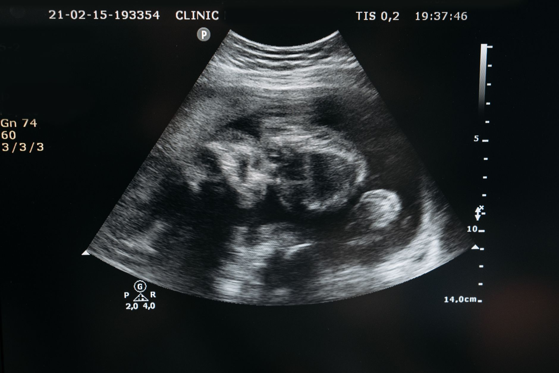
Typical ultrasound view of a cornular pregnancy: the sac sits outside the main uterine cavity.
What treatment options are available for a cornual pregnancy interstitial ectopic?
Management depends on gestational age, size of the gestational sac, patient stability, and desire for future fertility. The main approaches are:
Method
Typical Indication
Pros
Cons
Systemic methotrexate
Early (< 8 weeks), β‑hCG < 5000 mIU/mL, stable
Non‑surgical, preserves fertility, outpatient
May require multiple doses; slower resolution
Local methotrexate injection
Accessible sac, experienced operator
Higher drug concentration at target, less systemic exposure
Requires precise ultrasound guidance
Laparoscopic cornual resection
≥ 8 weeks, larger sac, or failed medical therapy
Immediate removal, definitive
Invasive, risk of uterine scarring
Robotic‑assisted cornular repair
Specialized centers, desire for precise suturing
Enhanced visualization, potentially less blood loss
Limited availability, higher cost
Hysterectomy (rare)
Massive hemorrhage, non‑viable uterus
Life‑saving when other measures fail
Loss of fertility
Medical therapy with methotrexate works by halting rapidly dividing trophoblastic tissue. It is most effective when the ectopic mass is smaller than 3.5 cm and the patient has no contraindications (e.g., liver disease, immunodeficiency).
Surgical options aim to excise the ectopic while preserving as much healthy uterine muscle as possible. Laparoscopic cornual resection involves a small incision, removal of the pregnancy tissue, and careful suturing of the uterine defect. The procedure carries a 10–15 % risk of postoperative bleeding, but most women return to normal activity within 4–6 weeks.
In emergency rupture, rapid laparotomy (open surgery) may be required to control hemorrhage. Blood transfusion and intensive monitoring are standard. Even in these urgent cases, surgeons strive to conserve the uterus when feasible.
Regardless of the method, follow‑up β‑hCG testing continues until levels are undetectable, confirming that all trophoblastic tissue has resolved. Some clinicians also schedule a repeat ultrasound 4–6 weeks after treatment to visualise scar healing.
What are the risks and complications of a cornual pregnancy interstitial ectopic?
The principal danger is sudden rupture leading to massive intra‑abdominal bleeding, which can cause hypovolemic shock. Reported mortality rates for ruptured interstitial ectopics range from 2–5 %—higher than for tubal ectopics—largely because the bleeding can be brisk and the diagnosis may be delayed.
Other complications include:
Uterine scarring that may affect future implantation or increase the risk of placenta accreta.
Reduced fertility if extensive myometrial tissue is removed or if adhesions form.
Persistent trophoblastic tissue that can cause a chronic ectopic or, rarely, a gestational trophoblastic disease.
Psychological distress—the loss of a pregnancy combined with the fear of surgery can trigger anxiety or depression.
Long‑term follow‑up includes monitoring for uterine integrity, especially if you plan another pregnancy. Many clinicians recommend waiting at least 3–6 months after surgery before attempting conception, though exact timing should be individualized.
Recent studies from the Fertility and Sterility journal (2022) suggest that women who undergo laparoscopic repair have a lower rate of subsequent uterine rupture compared with those who have a classical cornual resection, likely due to more precise suturing techniques.
How does a cornual pregnancy interstitial ectopic differ from a cesarean‑section scar pregnancy?
Both conditions involve implantation near or within a uterine scar, but they are distinct entities:
Location—A cornual pregnancy sits in the interstitial part of the tube within the uterine wall, whereas a cesarean‑section scar (C‑section) pregnancy implants directly into the fibrous scar tissue of a previous uterine incision.
Risk profile—C‑section scar pregnancies have a higher propensity for deep invasion into the myometrium and a greater chance of placenta accreta, while cornual pregnancies are more prone to early rupture due to limited muscular support.
Treatment—Both may be managed medically with methotrexate, but scar pregnancies often require combined approaches (e.g., uterine artery embolization plus curettage) because of their vascularity.
Imaging clues—On ultrasound, a scar pregnancy shows a gestational sac embedded within the scar with a “thin or absent” myometrial layer, whereas an interstitial ectopic shows the “interstitial line sign” and a sac located >1 cm from the uterine cavity.
The distinction matters because it guides the surgical plan and informs counseling about future pregnancy risks. If you have a history of cesarean delivery, your provider will pay special attention to the uterine scar during early scans to rule out both entities.
Guidelines from the NHS emphasize that scar pregnancies carry a higher risk of massive hemorrhage than cornual pregnancies, and therefore may warrant earlier intervention even when the sac is small.
What is the typical recovery time after surgery for a cornual pregnancy interstitial ectopic?
Recovery varies with the invasiveness of the procedure and the individual’s overall health. For minimally invasive laparoscopic resection, most patients report:
Hospital stay—1 day for uncomplicated cases; 2–3 days if blood loss was higher.
Return to light activities—Within 1–2 weeks, such as gentle walking or light housework.
Full return to work or exercise—Usually 4–6 weeks, depending on the physical demands of the job.
Sexual activity—Recommended to pause for at least 3 weeks or until the surgeon confirms healing via ultrasound.
Post‑operative pain is generally mild and manageable with acetaminophen or ibuprofen. Your provider will prescribe a short course of antibiotics to prevent infection and may suggest a pelvic binder for added support.
Emotional recovery is equally important. Many women experience grief over the loss of a pregnancy, coupled with anxiety about future fertility. Access to counseling, support groups, or a mental‑health professional can be beneficial. Keeping a journal of symptoms and emotions during the recovery weeks often helps identify lingering concerns early.
Physical therapists who specialize in postpartum care can also guide gentle core‑strengthening exercises once cleared by the surgeon, helping restore pelvic stability and reduce the risk of adhesions.
How should you track β‑hCG levels after treatment?
β‑hCG (beta‑human chorionic gonadotropin) is the hormone that confirms pregnancy. After medical or surgical treatment of a cornual ectopic, clinicians typically order serial β‑hCG measurements every 48–72 hours until the level drops below 5 mIU/mL. This pattern confirms that all trophoblastic tissue has been eliminated.
In practice, a rapid decline (greater than 50 % drop per interval) is reassuring, while a plateau or slow fall may signal retained tissue that could require additional methotrexate or a repeat surgical intervention. According to ACOG, failure to achieve a negative β‑hCG within 2–3 weeks warrants imaging to assess for persistent ectopic tissue.
Patients on methotrexate should also monitor for side effects such as mouth sores, liver enzyme changes, or bone‑marrow suppression. A baseline liver panel is usually obtained before treatment, and a repeat test is done a week after the dose. If any concerning symptoms arise—persistent fever, severe abdominal pain, or signs of infection—contact your provider promptly.
Tracking β‑hCG levels helps confirm that the ectopic tissue has resolved.
Lifestyle and reproductive planning to lower future cornual ectopic risk
While you cannot change a past scar, certain lifestyle choices can reduce the chance of another interstitial ectopic. Smoking cessation is paramount—cigarette smoke impairs tubal ciliary function, increasing the odds of abnormal embryo transport. The CDC reports that smokers have a 1.5‑fold higher risk of ectopic pregnancy overall.
Maintaining a healthy weight also matters. Obesity is linked to altered hormone levels that can affect tubal motility and implantation. A balanced diet rich in folate, iron, and omega‑3 fatty acids supports overall reproductive health and may improve uterine receptivity.
If you use an intrauterine device, discuss timing of removal with your provider when you’re ready to conceive. Some clinicians recommend a “wash‑out” period of one menstrual cycle after IUD removal before attempting conception, to allow the endometrium to normalize.
For couples undergoing IVF, careful embryo placement (avoiding the fundal region) and pre‑transfer hysteroscopy to assess scar integrity can lower the odds of cornual implantation. Many fertility centers now employ a “single‑embryo transfer” strategy to reduce the overall number of embryos that might implant in an abnormal location.
Fertility and future pregnancy after a cornual ectopic: what to expect
Most women who have successfully treated a cornual ectopic can achieve a healthy intrauterine pregnancy later on. A 2021 cohort study in *Obstetrics & Gynecology* followed 120 women after laparoscopic cornual resection; 86 % conceived within two years, and 78 % delivered a term baby without major complications.
Key factors that influence future fertility include the amount of myometrial tissue removed, the presence of adhesions, and the timing of the next conception. Your provider may recommend a follow‑up transvaginal ultrasound 3 months after surgery to confirm that the uterine wall has healed and that the scar thickness exceeds 5 mm—a threshold associated with lower risk of uterine rupture in subsequent pregnancies.
During a new pregnancy, early ultrasounds (6–8 weeks) are crucial to verify that the gestational sac is correctly positioned within the uterine cavity. Many obstetricians will schedule a “high‑resolution” scan at 12 weeks to assess scar integrity, especially if you had a prior cornual resection.
Psychologically, it’s normal to feel apprehensive about trying again. Couples often benefit from preconception counseling that addresses both medical and emotional concerns, helping to set realistic expectations and build confidence.
From our medical team: If you’re diagnosed with a cornual pregnancy, know that early detection dramatically improves outcomes. Most women can preserve their uterus and future fertility with either medical or minimally invasive surgical treatment. Always discuss the full range of options with a specialist, and don’t hesitate to ask about the impact on future pregnancies.
Myth vs. fact
Myth: Cornual pregnancies always require a hysterectomy. Fact: Only a small fraction—typically those with massive rupture—need hysterectomy. Most are treated with methotrexate or laparoscopic resection, preserving the uterus.
Myth: If you’ve had a cesarean, you’ll inevitably get a cornual pregnancy. Fact: Prior cesarean delivery increases risk but does not guarantee a cornual implantation. Proper early ultrasound can detect and manage it before complications arise.
Myth: You cannot become pregnant again after a cornual ectopic. Fact: Many women conceive successfully after appropriate healing. Follow‑up imaging and a recommended waiting period help optimize future pregnancy outcomes.
Key takeaways
Cornual (interstitial) pregnancy is a rare but high‑risk ectopic where the embryo implants in the uterine wall’s tubal segment.
Early warning signs include unilateral pelvic pain, spotting, and, in severe cases, shoulder pain or dizziness.
Risk factors include prior uterine surgery, IUD use, tubal abnormalities, and assisted reproductive technologies.
Diagnosis relies on transvaginal ultrasound, the “interstitial line sign,” and serial β‑hCG trends.
Treatment options range from systemic or local methotrexate to laparoscopic cornual resection; surgery is reserved for larger or ruptured pregnancies.
Recovery after minimally invasive surgery typically takes 4–6 weeks, with a gradual return to activity and close follow‑up.
Serial β‑hCG monitoring and scar‑healing ultrasounds are essential steps before attempting another pregnancy.
Lifestyle choices—like quitting smoking and maintaining a healthy weight—can lower the risk of recurrence.
Frequently asked questions
What are the chances of having a cornual pregnancy?
Cornual pregnancies account for less than 2 % of all ectopic pregnancies, which themselves occur in about 1–2 % of all conceptions. So the overall chance is roughly 0.02–0.04 % of all pregnancies.
Can you get pregnant again after a cornual pregnancy?
Yes. After the uterus has healed—usually 3–6 months—you can try to conceive again. Studies show that up to 80 % of women achieve a subsequent intrauterine pregnancy when the scar is intact and follow‑up imaging is normal.
How common is interstitial ectopic pregnancy?
Interstitial (cornual) ectopic pregnancy is the rarest form of ectopic, representing about 2–4 % of all ectopic pregnancies. It is therefore seen in roughly 1 in 5,000 pregnancies.
What are the symptoms of a ruptured ectopic pregnancy?
Sudden, severe abdominal or shoulder pain, faintness, rapid heartbeat, and signs of internal bleeding (pale skin, low blood pressure) indicate rupture and require emergency care.
Can a cornual pregnancy be carried to term?
In very rare cases, a cornual pregnancy has reached term, but the risk of uterine rupture and life‑threatening hemorrhage is extremely high. Current guidelines advise termination once the diagnosis is confirmed.
What are the risks of cornular pregnancy to the mother?
The main maternal risks are massive internal bleeding from rupture, need for blood transfusion, possible hysterectomy, and future uterine scarring that can affect fertility or increase obstetric complications.
Is methotrexate safe for breastfeeding after treatment?
Methotrexate is contraindicated during breastfeeding because the drug can pass into breast milk. The American College of Obstetricians and Gynecologists recommends waiting at least 24 hours after the final dose before resuming nursing, and many providers suggest pumping and discarding milk for a few days as an extra precaution.
Can a cornular pregnancy be detected on a routine early pregnancy scan?
Yes—if the scan is performed by a skilled sonographer at 6–8 weeks, the interstitial line sign can reveal a cornular implant. However, the rarity of the condition means it may be missed on a quick “first‑trimester” scan, so a dedicated “ectopic protocol” is advisable when risk factors are present.
When to call your doctor
If you experience sudden, severe abdominal or shoulder pain, heavy vaginal bleeding, dizziness, fainting, or a rapid heartbeat, seek emergency medical attention right away. For any persistent pelvic pain, spotting, or concerns about your pregnancy test results, contact your obstetrician or midwife promptly. This article provides general information only and is not a substitute for personalized medical advice.
References
American College of Obstetricians and Gynecologists (ACOG). “Management of Ectopic Pregnancy.” Practice Bulletin No. 193, 2020.
Royal College of Obstetricians and Gynaecologists (RCOG). “Ectopic Pregnancy.” Green‑top Guideline No. 1, 2021.
World Health Organization (WHO). “Recommendations for Prevention and Management of Ectopic Pregnancy.” 2022.
National Institute for Health and Care Excellence (NICE). “Ectopic Pregnancy: Diagnosis and Initial Management.” NG157, 2021.
Centers for Disease Control and Prevention (CDC). “Ectopic Pregnancy Surveillance.” 2023 data brief.
Mayo Clinic. “Cornual (Interstitial) Ectopic Pregnancy.” Updated 2023.
Fertility and Sterility Journal. “Outcomes After Medical vs. Surgical Treatment of Interstitial Ectopic Pregnancy.” 2022.
Obstetrics & Gynecology. “Robotic‑Assisted Cornular Resection: A Case Series.” 2021.
American College of Obstetricians and Gynecologists (ACOG). “Methotrexate Use in Pregnancy and Lactation.” Committee Opinion No. 752, 2020.
National Center for Biotechnology Information (NCBI). “Incidence of Interstitial Ectopic Pregnancy in IVF Cycles.” 2021.
British Medical Journal (BMJ). “Smoking and Ectopic Pregnancy Risk.” 2020.
Obstetrics & Gynecology. “Long‑Term Uterine Healing After Cornual Resection.” 2022.
Not sure about the label on Cornual Pregnancy Interstitial Ectopic Risks products?
Snap the ingredients list and SafeFilter checks every ingredient for your stage — only 3 free scans this month, then you're locked until reset. Unlimited from $7/mo or $50/yr.
When Shubhra Mishra was expecting her first child in 2016, she was overwhelmed by conflicting food advice — one site said yes, another said never. By the time her second baby arrived in 2019, she realized millions of mothers face the same confusion.
That sparked a five-year journey through clinical nutrition papers, cultural diets, and expert conversations — all leading to BumpBites: a calm, compassionate space where science meets everyday motherhood.
Her long-term vision is to build a global community ensuring safe, supported, and free deliveriesfor every mother — because no woman should face pregnancy alone or uninformed. 🌿
🌍 Stand with mothers, shape safer guidance
Join a small circle of experts who review BumpBites articles so expecting parents everywhere can decide with confidence.