Discover key development milestones visible in a 13 week pregnancy sonography. Learn what to expect, baby's growth, and important health insights for this stage.
By Shubhra Mishra — a mom of two who turned her own confusion during pregnancy into BumpBites, a global mission to make food choices clear, safe, and stress-free for every expecting mother. 💛
Check whether any food is safe during pregnancy with the BumpBites Food Safety Checker.
Quick take: At 13 weeks, an ultrasound reveals a fetus about the size of a lime, shows a steady heartbeat, and can capture the early shape of limbs, facial features, and the nuchal translucency space used for first‑trimester screening. Most measurements are within established ranges, making this scan a reliable tool for dating the pregnancy and spotting major anomalies.
It’s 2 a.m., you’ve just felt a flutter and are scrolling through pictures of “what a 13‑week baby looks like” while wondering whether the tiny heartbeat you heard on the monitor will keep going. You’re not alone—many expectant parents feel a mix of excitement and nerves before the first detailed scan of the second trimester.
In this guide we’ll walk through everything a 13‑week pregnancy sonography can show, from the numbers that tell you how far along you are to the tiny anatomical landmarks that signal healthy development. We’ll also cover what the scan feels like, how accurate it is for dating, what the nuchal translucency measurement means, and what to do if something looks off.
By the end you’ll know exactly what to expect during the appointment, which measurements are normal, how to interpret the heartbeat, and which early warning signs merit a quick call to your provider.
What does a 13‑week ultrasound show about fetal development?
At 13 weeks the fetus measures roughly 7–9 cm from crown to rump and weighs about 23 g—about the size of a lime or a small plum. This is the stage when the body starts to straighten out from the curled “bean” shape seen earlier, and the major organ systems are taking shape.
Key structures you’ll see on the screen include:
The head, now proportionally larger with a visible brain vesicle.
Two distinct ventricles in the brain, which appear as tiny fluid‑filled spaces.
The heart, beating at 110–160 beats per minute, often visualized in a four‑chamber view.
Developing limbs—upper arms, forearms, hands, thighs, and lower legs—with tiny fingers and toes beginning to form.
Facial features such as the upper lip, nose bridge, and the beginnings of the eyelids.
The nuchal translucency (NT) space, a fluid‑filled pocket at the back of the neck that is measured for screening of chromosomal conditions.
While the fetus is still too small for detailed anatomy that will be done later in the second trimester, these landmarks give a reassuring snapshot of overall growth.
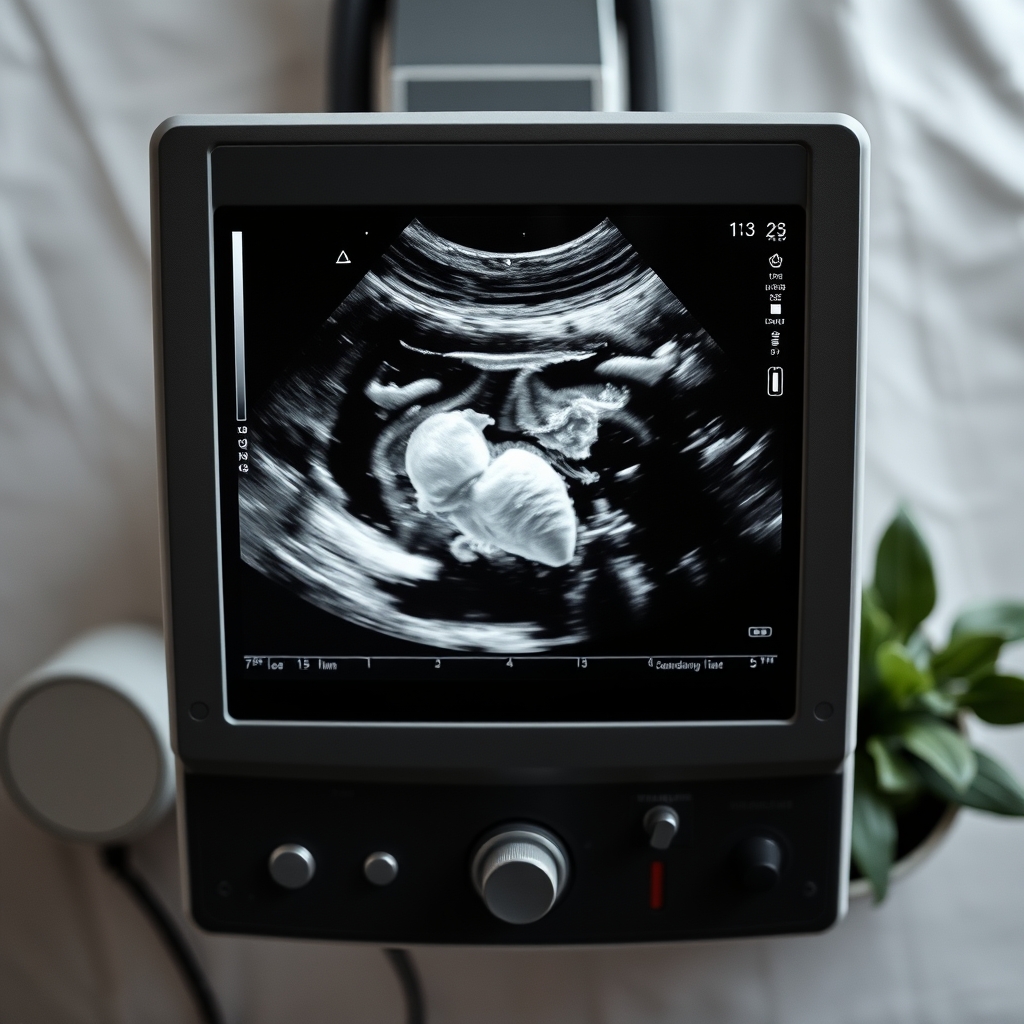
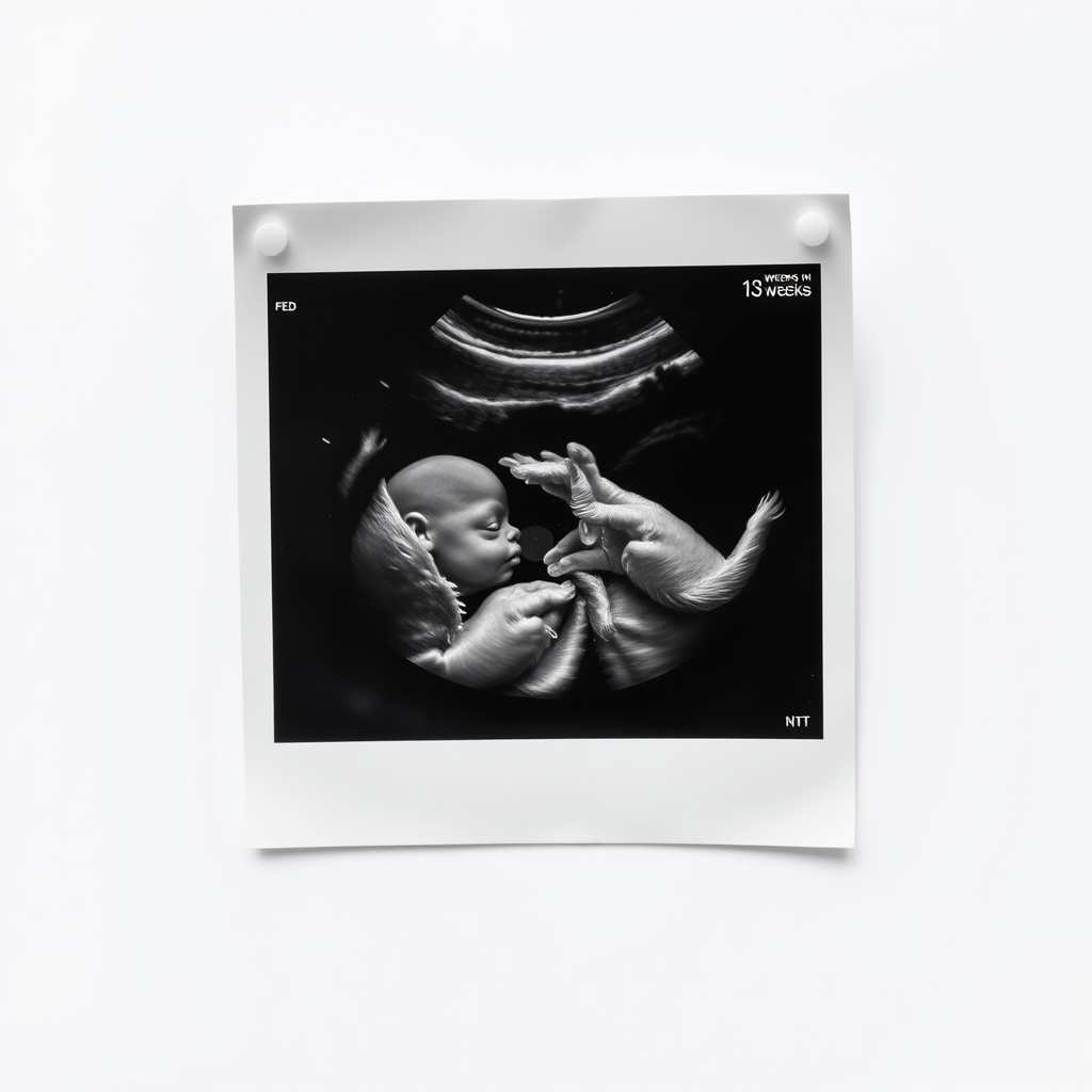
Most parents ask to see pictures of a 13‑week scan. Below is a placeholder for a typical image that will appear in the final article.
A typical 13‑week ultrasound image displaying early limb and facial development.
Beyond the structures listed, the sonographer also evaluates the overall shape of the fetal torso and the position of the placenta. A well‑positioned placenta at this stage is a good sign, although the exact location (anterior vs. posterior) won’t be definitively classified until later scans. Seeing the fetus in a relaxed, neutral position helps ensure that measurements are not skewed by flexion or compression.
What measurements does a 13‑week sonogram include and what do they mean?
The sonographer will take several standard measurements that help confirm gestational age and screen for potential issues. The most common are:
Crown‑Rump Length (CRL) – distance from the top of the head to the bottom of the torso.
Head Circumference (HC) – the perimeter of the skull, useful for tracking brain growth.
Femur Length (FL) – length of the longest bone in the body, indicating limb development.
Nuchal Translucency (NT) thickness – the fluid space behind the neck, part of the combined first‑trimester screen.
Below is a quick reference table of normal ranges for a singleton pregnancy at 13 weeks. Values outside these ranges don’t automatically mean a problem, but they prompt a closer look.
Measurement
Normal range (13 weeks)
What it indicates
Crown‑Rump Length (CRL)
7.0–9.0 cm
Confirms gestational age; very small CRL may suggest earlier dating error.
Head Circumference (HC)
16.0–22.0 mm
Assesses early brain growth; markedly low HC can signal microcephaly.
Femur Length (FL)
6.5–9.0 mm
Tracks limb development; shortened FL may hint at skeletal dysplasia.
Nuchal Translucency (NT)
≤ 3.5 mm (adjusted for CRL)
Elevated NT can be associated with chromosomal abnormalities, especially when combined with blood markers.
These numbers are obtained quickly—usually within the first 10 minutes of the scan—and are compared against standardized charts from the World Health Organization (WHO) and the American College of Obstetricians and Gynecologists (ACOG). The more closely the measurements align with expected values, the more confidence you and your provider have in the dating and early screening results.
In addition to the core measurements, the sonographer may note the position of the fetal spine (a straight line suggests normal development) and the presence of any extra‑uterine fluid collections, which can occasionally signal early miscarriage risk. All of these observations are documented in the scan report for your provider’s review.
How do I interpret the fetal heartbeat on a 13‑week ultrasound?
The heartbeat is one of the most reassuring features you’ll see. At 13 weeks the heart typically beats between 110 and 160 times per minute. The sonographer will usually capture a Doppler audio clip, letting you hear the “whoosh‑whoosh” rhythm for yourself.
If the heart rate falls below 110 bpm or exceeds 160 bpm, the sonographer may repeat the measurement after a short break. A slightly slower rate can be normal if the fetus is briefly resting, while an unusually rapid rate may be linked to fever or maternal anxiety.
Beyond the rate, the four‑chamber view of the heart is examined for structural integrity. At this stage, major heart defects such as atrial septal defects are still too small to diagnose definitively, but gross abnormalities (e.g., single ventricle) can sometimes be spotted.
Another interesting milestone is fetal movement. By 13 weeks, the baby begins to make spontaneous, though still subtle, motions that may be visible as tiny twitches on the screen. These movements are a good sign of neuromuscular development and often precede the first maternal perception of “quickening,” which usually occurs a few weeks later.
It’s also worth noting that maternal factors—such as caffeine intake or recent exercise—can transiently affect fetal heart rate. If your provider sees a borderline reading, they may ask about recent activities and possibly schedule a repeat scan to confirm stability.
Can you see the baby’s limbs and facial features at 13 weeks?
Yes—by the middle of the first trimester the limbs are long enough to be distinguished, and the face is taking on recognizable features. You’ll typically see:
Upper and lower arms with elbows and wrists, plus tiny fingers beginning to separate.
Thighs and lower legs with a visible knee joint; the toes are still webbed but the outline is clear.
The upper lip, nose bridge, and the beginnings of the eyelids, though the eyes are still closed.
In most cases, the gender cannot be determined reliably at 13 weeks; the genital tubercle is still forming.
Because the skull is still relatively large compared to the body, the facial profile may appear slightly flattened. The sonographer may angle the probe to capture a “profile” view that highlights the nose and lip contour.
While limb and facial visibility is reassuring, it’s also a chance to check for major malformations such as limb‑body wall complex or severe facial clefts. These are rare, but early detection allows for timely counseling and planning.
Occasionally, the sonographer can capture a brief “hand‑wave” motion, where the fetus opens and closes its tiny fingers. This subtle movement is an early sign of developing voluntary motor control and is often a source of delight for parents watching the screen.
What is a normal crown‑rump length range for a 13‑week gestation ultrasound?
The CRL is the most reliable single measurement for dating a pregnancy in the first trimester. At 13 weeks, the expected CRL is between 7.0 cm and 9.0 cm (approximately 2.8–3.5 inches). Measurements slightly outside this window can still be normal, especially if the ovulation date is uncertain.
When the CRL is smaller than 7 cm, clinicians often review the dating based on the last menstrual period (LMP) and may repeat the scan in a week to see if growth catches up. Conversely, a CRL larger than 9 cm may suggest a later gestational age, prompting a re‑evaluation of the due date.
In practice, the sonographer will also record HC, FL, and NT to create a composite picture. Consistency across multiple measurements increases confidence in the gestational age estimate.
It’s helpful to remember that a small variation—say, a CRL of 6.9 cm—does not automatically indicate a problem. The fetus can grow at slightly different rates, and a single measurement is only one piece of the overall picture.
What abnormalities can be detected during a 13‑week sonography?
At 13 weeks the scan is primarily a screening tool rather than a definitive diagnostic exam. However, certain major anomalies can be identified, including:
Severe neural tube defects (e.g., anencephaly) that present as an absent skull vault.
Large abdominal wall defects such as omphalocele, where the intestines protrude into the base of the umbilical cord.
Major cardiac malformations that alter the four‑chamber view.
Chromosomal risk markers like increased nuchal translucency thickness.
Early signs of multiple gestation (e.g., two distinct sacs) that may impact management.
The combined first‑trimester screen—NT measurement plus maternal serum markers (PAPP‑A and free β‑hCG)—is the most sensitive way to assess the risk for Down syndrome and other chromosomal conditions at this stage. A high NT measurement alone does not confirm a problem but does raise the calculated risk, prompting further testing such as non‑invasive prenatal testing (NIPT) or a detailed anatomy scan later in the second trimester.
If an abnormality is suspected, the sonographer will usually recommend a follow‑up targeted scan at 18–22 weeks for a more thorough evaluation. In some cases, a referral to a fetal medicine specialist may be advised earlier.
Some rare conditions, such as severe congenital diaphragmatic hernia, can also be hinted at by abnormal fluid collections or unusual organ positioning. While these findings are uncommon at 13 weeks, early identification enables multidisciplinary planning and, when appropriate, timely intervention.
How accurate is the 13‑week ultrasound for dating the pregnancy?
Dating accuracy is highest between 8 and 14 weeks because fetal growth is relatively uniform. Studies referenced by the Royal College of Obstetricians and Gynaecologists (RCOG) show that a CRL measurement at 13 weeks predicts the actual date of delivery within ± 5 days in about 80 % of cases.
This is more precise than measurements taken later in the second trimester, where variations in fetal size increase due to genetics, maternal health, and nutrition. Therefore, if you haven’t had an early scan (9–11 weeks), the 13‑week scan becomes the benchmark for establishing your estimated due date (EDD).
When a discrepancy between the LMP‑based due date and the CRL‑based estimate is noted, clinicians will usually adopt the CRL date, especially if the difference exceeds 7 days. This helps align prenatal care milestones such as glucose screening and anatomy scans with the most accurate gestational age.
It’s also worth noting that ultrasound dating remains reliable even in women with higher body mass index (BMI); the quality of the image may be slightly reduced, but the CRL measurement is still considered the gold standard for dating in the first trimester, according to ACOG guidelines.
What is the purpose of nuchal translucency screening at 13 weeks?
The nuchal translucency (NT) measurement is a key component of the combined first‑trimester screen. A fluid‑filled space behind the fetal neck is measured in millimeters; the larger the NT, the higher the risk for chromosomal abnormalities like trisomy 21 (Down syndrome) and trisomy 18.
Guidelines from ACOG and the National Health Service (NHS) recommend measuring NT when the CRL is between 45 mm and 84 mm—exactly the range you’ll encounter at 13 weeks. The measurement is taken in a sagittal plane with the fetus’s head and spine aligned, and the sonographer will often capture a still image for later review.
NT alone is not diagnostic. It is combined with maternal serum markers (PAPP‑A and free β‑hCG) and maternal age to calculate a risk score. A low risk (e.g., < 1:1000) is reassuring, while a higher risk may lead to counseling about options such as cell‑free DNA testing (NIPT) or a diagnostic chorionic villus sampling (CVS) if you’re comfortable with invasive testing.
Because NT measurement is technically sensitive, many clinics follow a strict quality‑control protocol, including double‑reading by a second sonographer and adherence to the 2016 International Society of Ultrasound in Obstetrics and Gynecology (ISUOG) standards.
NT measurement is taken when the CRL is 45–84 mm, typically during the 13‑week scan.
What can you expect during a 13‑week prenatal scan appointment?
The appointment usually lasts 20–30 minutes. After a brief check of your medical history, you’ll lie on an exam table while a gel is applied to your abdomen. The sonographer slides a transducer over your skin, capturing images in real time.
Typical steps include:
Confirming gestational age with CRL.
Measuring head circumference, femur length, and NT.
Assessing the heart rate and four‑chamber view.
Visualizing limbs, facial profile, and overall fetal position.
Printing or sending a digital report with key measurements and any notable observations.
Most women report feeling warm from the gel, a gentle pressure from the probe, and a sense of reassurance when they hear the heartbeat. The scan is safe—no radiation, and the FDA classifies diagnostic ultrasound as a Category II device, meaning it’s considered safe when used appropriately.
During the exam, the sonographer may also ask you to take a deep breath or to gently shift position to get the best view of the fetal spine and placenta. These small adjustments help produce clearer images without prolonging the visit.
Early signs of miscarriage on a 13‑week ultrasound
While most 13‑week scans are normal, certain findings may suggest a non‑viable pregnancy:
No detectable heartbeat after repeated attempts.
Irregular or absent fetal cardiac activity.
Markedly abnormal CRL for the gestational age (e.g., < 6 cm at 13 weeks).
Presence of a “blighted ovum” where a gestational sac is seen without an embryo.
If any of these are observed, your provider will discuss next steps, which may include a repeat scan in 7–10 days, blood hCG monitoring, or, if confirmed, appropriate counseling and care options.
Differences between 12‑week and 13‑week ultrasounds
Both scans aim to confirm dating and screen for early anomalies, but the 13‑week scan offers a few advantages:
Greater limb length makes elbows, wrists, and finger buds easier to see.
The NT measurement is more reliable because the CRL falls squarely within the recommended window.
Fetal cardiac structures are larger, improving the chances of obtaining a clear four‑chamber view.
Overall image clarity is higher, reducing the need for repeat scanning.
That said, a 12‑week scan can still capture many of the same landmarks, especially if the fetus is well positioned. If you missed a 12‑week appointment, the 13‑week scan is an excellent backup.
13‑week gestational age ultrasound checklist
Here’s a quick reference you can bring to your appointment:
Last menstrual period (LMP) date and any known ovulation timing.
Previous ultrasound reports (if any) for comparison.
List of questions about NT, fetal heartbeat, and any personal risk factors.
Contact information for your obstetric provider in case you need follow‑up.
Having these items handy helps the sonographer focus on the most relevant measurements and ensures you leave with the information you need.
How to prepare for a 13‑week ultrasound
Preparation is simple, but a few small steps can improve image quality and reduce anxiety. Arrive with a full bladder if your provider asks for it—this lifts the uterus higher into the abdomen, giving the transducer a clearer window. Drink about 500 ml of water 30 minutes before your appointment and avoid emptying your bladder until after the scan.
If you’re feeling nauseous, a light snack (such as a piece of toast with a thin spread of almond butter) can settle your stomach without causing excess gas, which can interfere with imaging. Dress in loose‑fitting clothing and wear a shirt that can be easily lifted or slid up, so the sonographer can access your abdomen without hassle.
Mentally, give yourself a few minutes before the visit to breathe deeply and visualize a calm environment. Many clinics play soft music or have a quiet room, but bringing headphones with your favorite calming playlist can help you stay relaxed while the images are captured.
Nutrition and hydration tips for optimal imaging
Staying well‑hydrated not only helps with a full bladder but also supports overall fetal health. Aim for 8–10 glasses of water per day, and consider adding a pinch of sea salt to your water if you’re prone to dehydration. A balanced snack that includes protein (e.g., a hard‑boiled egg) and a small amount of complex carbohydrate (whole‑grain crackers) can keep blood sugar stable, reducing the chance of sudden fetal movement that might blur the picture.
Here’s a quick reference table of foods that are both safe and beneficial before your scan:
Food
Why it helps
Portion
Greek yogurt
Provides protein and calcium for muscle tone
½ cup
Banana
Potassium supports fluid balance
1 small
Whole‑grain toast
Complex carbs keep blood sugar steady
1 slice
These options are gentle on the stomach and unlikely to cause excess gas, which can obscure the ultrasound view. Avoid heavy, fried, or very spicy foods right before the appointment, as they may increase gastrointestinal discomfort.
What are the next steps if the nuchal translucency measurement is high?
A higher‑than‑average NT reading can be unsettling, but it’s only one piece of the risk puzzle. Your provider will first review the NT measurement alongside the CRL to ensure it’s correctly adjusted. If the NT remains elevated, the combined first‑trimester screen will be recalculated with your maternal serum markers.
Depending on the calculated risk, you may be offered non‑invasive prenatal testing (NIPT), which analyzes fetal DNA fragments in maternal blood and has a detection rate above 99 % for common trisomies. If you prefer a definitive diagnosis, chorionic villus sampling (CVS) or amniocentesis can be discussed, though these are invasive and carry a small risk of miscarriage.
Regardless of the path you choose, most families with an elevated NT result go on to have healthy pregnancies. The key is early, open communication with your obstetric team, who can guide you through the options and support your decision‑making process.
From our medical team: A 13‑week scan is both a milestone and a safety net. It confirms that your baby is growing as expected, provides a reliable due‑date anchor, and offers the first glimpse at the heart and neck that can flag higher‑risk conditions. If any measurement falls outside the typical range, remember that most variations are benign and can be clarified with a follow‑up scan. Always discuss any concerns with your obstetrician or midwife—they’ll tailor the next steps to your unique situation.
Myth vs. fact
Myth: You can determine the baby’s gender at 13 weeks. Fact: The genital tubercle is still forming, so gender identification is unreliable until around 18–20 weeks.
Myth: An ultrasound can cause harm to the fetus. Fact: Diagnostic ultrasound uses sound waves, not radiation, and is considered safe when performed by trained professionals (FDA, ACOG).
Myth: A normal 13‑week scan guarantees a complication‑free pregnancy. Fact: While reassuring, the scan only assesses early development; later anomalies can still emerge, so continued prenatal care remains essential.
Key takeaways
At 13 weeks the fetus is 7–9 cm long, with a heartbeat of 110–160 bpm and visible limbs, facial features, and nuchal translucency.
Standard measurements (CRL, HC, FL, NT) have defined normal ranges; deviations prompt closer monitoring.
The scan is the most accurate dating tool in the first trimester, usually within ± 5 days of the true due date.
Nuchal translucency screening, combined with maternal serum markers, assesses early risk for chromosomal conditions.
The procedure is safe, painless, and typically lasts 20–30 minutes; bring a checklist of your LMP and any prior results.
Red‑flag signs—no heartbeat, absent embryo, or markedly abnormal measurements—should prompt a follow‑up scan or a call to your provider.
Frequently asked questions
What can you see on a 13‑week ultrasound?
Immediately you’ll see the fetal heartbeat, a clear crown‑rump length, the four‑chamber view of the heart, developing limbs, and early facial features such as the upper lip and nose bridge, plus the nuchal translucency space.
Is a 13‑week ultrasound safe for the baby?
Yes. Diagnostic ultrasound uses low‑energy sound waves, not ionizing radiation, and is classified by the FDA as safe when performed by a qualified professional.
How long does a 13‑week prenatal scan take?
Most appointments last 20–30 minutes, including set‑up, measurement collection, and a brief review of findings with the sonographer.
What are the normal measurements at 13 weeks?
Typical values are a CRL of 7.0–9.0 cm, head circumference of 16–22 mm, femur length of 6.5–9 mm, and NT thickness ≤ 3.5 mm (adjusted for CRL).
Can you see the baby’s gender at 13 weeks?
Gender is usually not discernible at this stage because the genital tubercle is still developing; most providers wait until 18–20 weeks for a reliable determination.
What abnormalities are checked at a 13‑week ultrasound?
The scan screens for major neural tube defects, large abdominal wall defects, significant heart malformations, and elevated nuchal translucency that may indicate chromosomal risks.
Will a higher‑than‑average nuchal translucency affect my pregnancy plan?
A high NT reading prompts a reassessment of risk using maternal serum markers and may lead to offering non‑invasive prenatal testing (NIPT) or, if desired, diagnostic testing like CVS. Most families with an elevated NT continue to have healthy pregnancies, and your care team will guide you through the options.
Can a woman with a higher BMI still get a clear 13‑week scan?
Yes. While increased abdominal tissue can slightly reduce image clarity, the CRL measurement remains reliable for dating. Technologists may adjust the probe pressure or use higher‑frequency settings to obtain the best possible view, as recommended by ACOG.
When to call your doctor
If the sonographer reports any of the following, contact your obstetric provider right away: no detectable heartbeat after repeated attempts, a CRL significantly smaller or larger than expected for 13 weeks, elevated nuchal translucency without a clear explanation, or any mention of a structural abnormality.
This article is for informational purposes only and does not replace personalized medical advice. Always discuss your specific situation with a qualified health professional.
References
American College of Obstetricians and Gynecologists (ACOG). “Ultrasound Guidance for Pregnancy Dating.” Practice Bulletin No. 171, 2016.
World Health Organization (WHO). “Guidelines for Ultrasound in Pregnancy.” 2020.
Royal College of Obstetricians and Gynaecologists (RCOG). “Nuchal Translucency Scan.” Green‑top Guideline No. 57, 2021.
National Health Service (NHS). “First Trimester Screening.” Updated 2022.
International Society of Ultrasound in Obstetrics and Gynecology (ISUOG). “Guidelines for the Performance of the Nuchal Translucency Scan.” 2016.
U.S. Food and Drug Administration (FDA). “Diagnostic Ultrasound Safety.” 2023.
Not sure about the label on 13 Week Pregnancy Sonography Development Milestones products?
Snap the ingredients list and SafeFilter checks every ingredient for your stage — only 3 free scans this month, then you're locked until reset. Unlimited from $7/mo or $50/yr.
When Shubhra Mishra was expecting her first child in 2016, she was overwhelmed by conflicting food advice — one site said yes, another said never. By the time her second baby arrived in 2019, she realized millions of mothers face the same confusion.
That sparked a five-year journey through clinical nutrition papers, cultural diets, and expert conversations — all leading to BumpBites: a calm, compassionate space where science meets everyday motherhood.
Her long-term vision is to build a global community ensuring safe, supported, and free deliveriesfor every mother — because no woman should face pregnancy alone or uninformed. 🌿
🌍 Stand with mothers, shape safer guidance
Join a small circle of experts who review BumpBites articles so expecting parents everywhere can decide with confidence.