Safe: Dental Xray is low‑risk during pregnancy when exposure stays below 0.01 mSv, particularly after the first trimester. Find dosage limits, trimester guidelines, and safer alternatives.
By Shubhra Mishra — a mom of two who turned her own confusion during pregnancy into BumpBites, a global mission to make food choices clear, safe, and stress-free for every expecting mother. 💛
Check whether any food is safe during pregnancy with the BumpBites Food Safety Checker.
Quick verdict: ⚠️ Talk to your doctor first. Dental X‑rays are generally considered low‑risk, but you should only have them when necessary and with proper shielding.
It’s completely normal to feel a flutter of anxiety the moment you hear “X‑ray” while you’re pregnant. Whether you’re in the middle of a routine dental check‑up or you’ve just been told a bite‑wing is needed, the question that pops up is “is dental X‑ray safe during pregnancy?” The short answer is that most dental X‑rays deliver a very small amount of radiation, and with lead shielding they are usually safe, but they should be used judiciously and only when the benefit outweighs any theoretical risk.
In this article we’ll walk you through exactly what a dental X‑ray is, what the current guidance from the American College of Obstetricians and Gynecologists (ACOG), the NHS, and the FDA says, and how safety changes across each trimester. We’ll also cover how many X‑rays are considered acceptable, what precautions you and your dentist can take, and what safer imaging alternatives exist if you’d rather avoid radiation altogether. By the end you’ll have a clear, evidence‑based roadmap to help you discuss any concerns with your provider and stop worrying.
Many expectant parents wonder whether a routine bite‑wing taken at a six‑month check‑up could affect their baby’s development. Others worry that postponing an X‑ray might let a cavity worsen, potentially leading to infection that could harm both mother and child. We’ll address both sides of that coin, explain the real numbers behind radiation exposure, and give you practical tips for talking with your dentist and obstetrician so you can feel confident in the care you receive.
Stage of pregnancy
Safety verdict
Notes
First trimester
⚠️ Use only if essential
Lead apron recommended; limit to urgent cases
Second trimester
✅ Generally safe with shielding
Routine exams usually permissible
Third trimester
✅ Generally safe with shielding
Same precautions as second trimester
Breastfeeding
✅ No additional risk
Radiation does not pass into breast milk
What is a dental X‑ray?
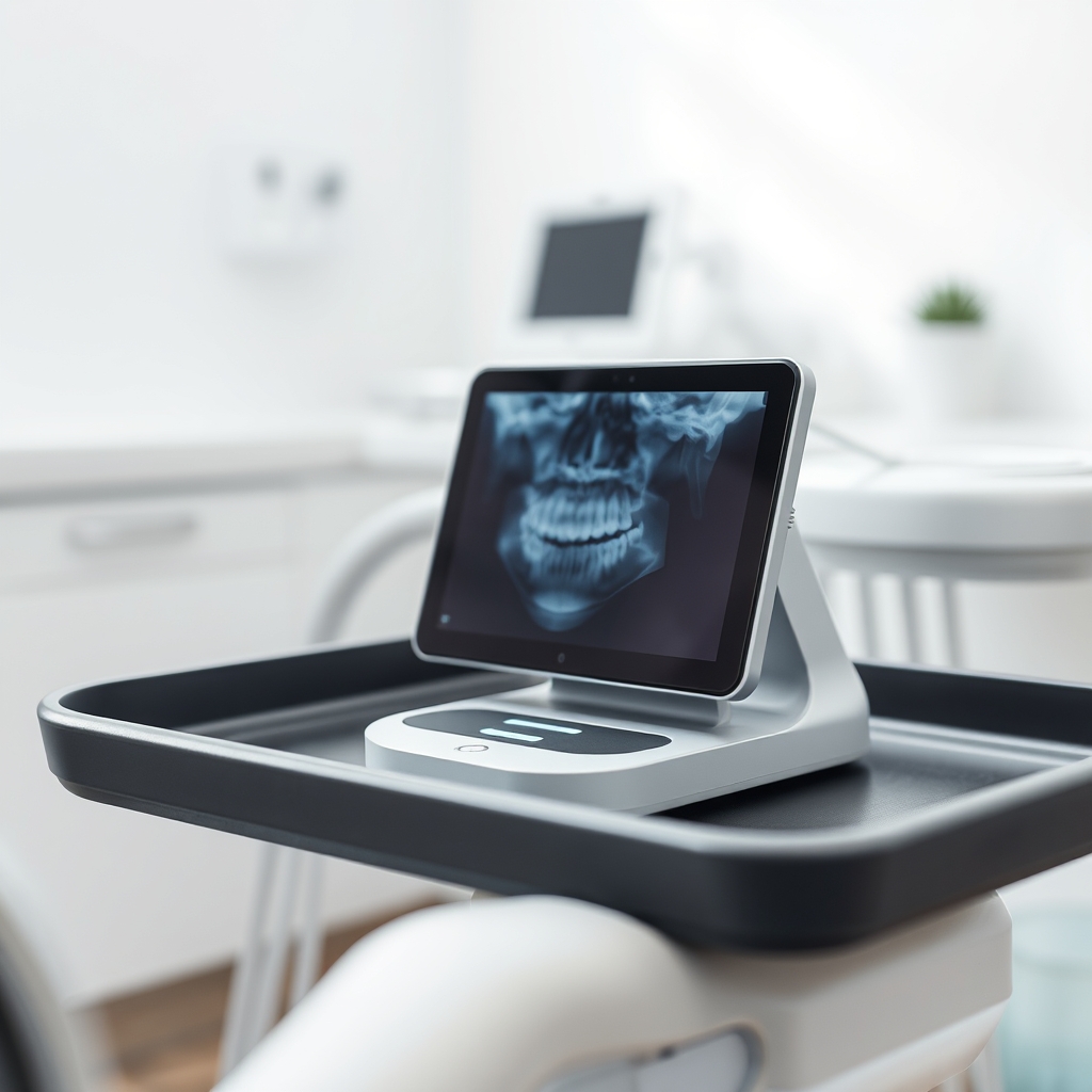
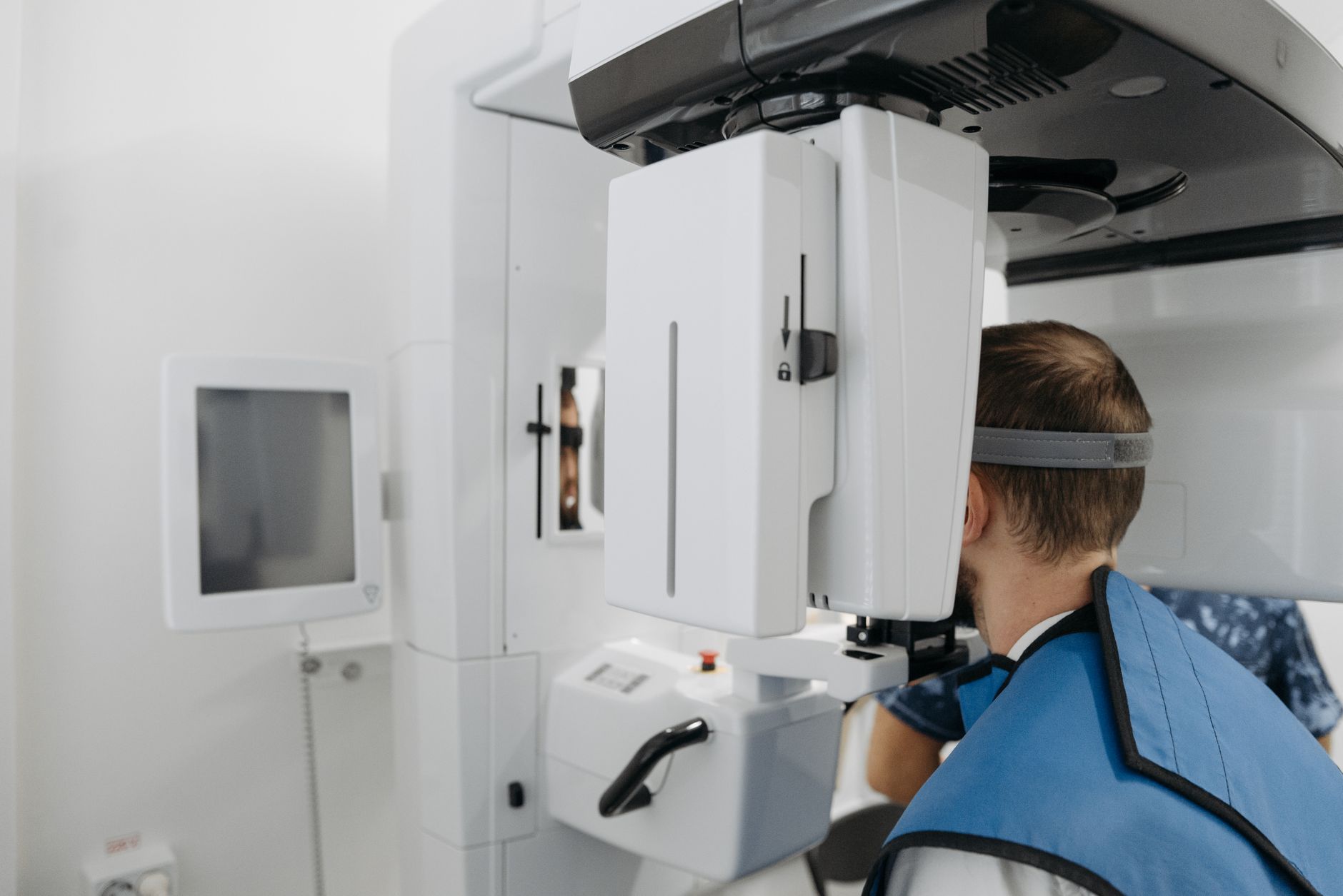
A dental X‑ray is a small, focused burst of ionising radiation that creates an image of your teeth, jawbone, and surrounding structures. The most common types are bite‑wing X‑rays (which show the crowns of the upper and lower teeth), periapical X‑rays (which capture the whole tooth from crown to root), and panoramic X‑rays (which provide a broad view of the entire mouth in a single image). Modern dental offices typically use digital intraoral radiography, which reduces exposure compared with older film‑based systems. Dentists order X‑rays to detect cavities, assess bone loss, plan orthodontic treatment, or evaluate the health of dental implants.
Because the radiation is directed only at the mouth, the dose is far lower than that of a chest X‑ray or a CT scan. Nevertheless, any exposure to ionising radiation during pregnancy raises questions about potential effects on a developing fetus, which is why the safety of dental X‑rays is a common concern among expectant parents.
Digital sensors have largely replaced traditional film, meaning the X‑ray beam can be turned off as soon as the image is captured, cutting exposure time by up to 70 %. Some offices also use extra‑low‑dose settings specifically for pregnant patients, further shrinking the already tiny dose.
Is dental X‑ray safe during pregnancy?
Current guidance from leading health organisations indicates that a single dental X‑ray, when performed with proper lead shielding, delivers an amount of radiation that is well below the threshold associated with fetal harm. The American College of Obstetricians and Gynecologists (ACOG) states that “the fetal radiation dose from a typical intraoral dental X‑ray is negligible” and that routine dental care should not be postponed if it is needed for oral health. The UK’s National Health Service (NHS) echoes this sentiment, noting that dental X‑rays are safe when a lead apron is used and that the benefit of treating a dental infection outweighs any minimal risk.
The U.S. Food and Drug Administration (FDA) classifies dental X‑ray devices as low‑dose radiation sources. According to the FDA, the effective dose from a single intraoral X‑ray is about 0.005 millisieverts (mSv), which is roughly 1/1000th of the average background radiation a person receives in a year. The Centers for Disease Control and Prevention (CDC) adds that cumulative exposure should stay below the 5 mSv limit recommended for the general public during pregnancy, a level far higher than what would be accumulated from a few dental X‑rays.
Evidence from epidemiological studies shows no statistically significant increase in birth defects, miscarriage, or developmental delays linked to dental X‑ray exposure when proper shielding is used. A large cohort study from the 1990s that followed over 2,000 pregnant women found that those who had dental X‑rays reported no higher rates of adverse outcomes compared with unexposed controls.
In short, the evidence suggests that a dental X‑ray is safe during pregnancy when necessary, especially if your dentist uses a lead apron and follows standard radiation‑reduction protocols. However, because the first trimester is a period of rapid organ formation (organogenesis), many clinicians prefer to limit exposure to only urgent cases during that time.
Is a dental X‑ray safe in the first trimester of pregnancy?
During the first trimester, the fetus is most vulnerable to teratogens—agents that could cause birth defects—because its organs are forming. While the radiation dose from a dental X‑ray is still extremely low, most obstetric guidelines advise that X‑rays be reserved for urgent dental issues (such as severe infection or trauma) during this period. If a dental X‑ray is deemed essential, a lead apron covering the abdomen and thyroid should be used, and the dentist should employ the lowest‑possible exposure settings.
ACOG notes that “if a dental X‑ray is unavoidable, the benefit to the mother’s oral health generally outweighs the minimal fetal risk.” In practice, many dentists will postpone routine imaging until the second trimester, when the risk of radiation‑induced teratogenic effects is considerably lower.
Some practitioners also recommend using a thyroid collar in addition to the abdominal lead apron, because the thyroid gland is particularly sensitive to radiation. This extra precaution adds virtually no inconvenience but further reduces any theoretical risk.
How many dental X‑rays are safe during pregnancy?
There is no official “hard limit” on the number of dental X‑rays a pregnant person can have, but the cumulative dose should stay well under the 5 mSv threshold set by the FDA for the general public. A typical bite‑wing X‑ray delivers about 0.005 mSv, meaning you could theoretically have up to 1,000 bite‑wing images before reaching that limit—far more than any realistic dental treatment would require.
In practical terms, most dental professionals limit exposure to one or two bite‑wing or periapical X‑rays per appointment, and they will only repeat imaging if a significant change in the oral condition occurs. If you need multiple images (for example, a full‑mouth series), your dentist will usually space them out and ensure each image is justified.
When a full‑mouth series is required, the total dose typically stays under 0.05 mSv, which is still less than 1 % of the background radiation you receive in a year. Even in the unlikely event of needing several series over the course of a pregnancy, the cumulative exposure remains well below safety limits.
What are the risks of dental X‑rays for pregnant women?
The primary risk associated with any ionising radiation is the theoretical chance of DNA damage that could affect fetal development. However, the dose from a dental X‑ray is so low that the risk is considered negligible. Studies have not demonstrated a statistically significant increase in birth defects, miscarriage, or developmental delays linked to dental X‑ray exposure when proper shielding is used.
Other, more immediate concerns involve the mother: excessive radiation without shielding could cause skin erythema or, very rarely, thyroid irritation. That’s why the use of a lead apron and thyroid collar is standard practice. For the fetus, the only measurable risk is a minute increase in the probability of a minor anomaly, which is far outweighed by the benefits of treating dental infections that could otherwise lead to systemic inflammation.
It’s also worth noting that untreated dental disease itself can pose a risk to pregnancy. Severe periodontal disease has been associated with preterm birth and low birth weight, so timely diagnosis and treatment—sometimes requiring an X‑ray—can actually protect both mother and baby.
Are there safer alternatives to dental X‑rays for pregnant patients?
When radiation exposure is a concern, several non‑ionising imaging methods can provide valuable diagnostic information:
Digital intraoral radiography – uses lower doses than traditional film.
Panoramic dental X‑ray with lead apron – offers a broader view while still keeping exposure minimal.
Ultrasound dental imaging – emerging technology that visualises soft‑tissue structures without radiation.
Clinical visual dental examination – relies on visual inspection and probing, sufficient for many routine checks.
Photographic dental impressions – high‑resolution photos can help monitor changes over time without radiation.
MRI dental imaging – provides detailed images of soft tissues without ionising radiation, though not commonly used for routine dental diagnostics.
These alternatives are especially useful for monitoring existing conditions or planning non‑urgent procedures. Discuss them with your dentist to see if any fit your specific situation.
Do dental X‑ray brands differ in radiation safety for pregnant women?
Modern dental X‑ray units, whether from manufacturers such as Sirona, Carestream, or Planmeca, all adhere to strict safety standards set by the International Electrotechnical Commission (IEC) and the FDA. The differences between brands largely involve image quality, speed, and ergonomic design rather than radiation dose. All reputable brands incorporate dose‑reduction technologies like rectangular collimation and digital sensors, which lower exposure compared with older, film‑based systems.
When choosing a dental practice, ask whether they use digital intraoral radiography and whether they provide a lead apron for pregnant patients. The specific brand of the X‑ray machine is less important than the use of proper shielding and low‑dose protocols.
Can dental X‑rays cause birth defects?
Current evidence does not support a causal link between dental X‑rays and birth defects. The fetal dose from a single intraoral X‑ray is far below the threshold associated with teratogenic effects. ACOG and the NHS both state that, with appropriate lead shielding, the risk of causing a birth defect is essentially negligible. However, if a dental X‑ray is performed without shielding or in the context of multiple unnecessary exposures, the theoretical risk could increase, which is why precautionary measures are emphasized.
What precautions should be taken for dental X‑rays during pregnancy?
To minimise any potential risk, the following steps are recommended:
Inform your dentist of your pregnancy status as early as possible.
Request a lead apron that covers the abdomen and thyroid.
Ask that the dentist use the lowest effective exposure setting and rectangular collimation.
Limit imaging to the smallest area necessary (e.g., bite‑wing rather than full‑mouth series) unless a broader view is essential.
Schedule routine dental X‑rays for the second trimester when possible.
Discuss any concerns with your obstetric provider, especially if you have a high‑risk pregnancy.
How does a dental X‑ray dosage compare to other medical imaging during pregnancy?
In the spectrum of medical imaging, dental X‑rays sit at the very low‑dose end. For perspective:
Dental bite‑wing X‑ray: ~0.005 mSv
Chest X‑ray: ~0.02 mSv
Abdominal X‑ray: ~0.7 mSv
CT scan of the head: ~2 mSv
CT scan of the abdomen/pelvis: ~10 mSv
All of these figures are approximate and can vary based on equipment and technique. Even the higher‑dose CT scans remain below the 5 mSv limit for the general public, but they are still substantially greater than a dental X‑ray. The key takeaway is that a dental X‑ray’s radiation dose is minuscule compared with most other imaging studies performed during pregnancy.
Modern digital dental X‑ray sensors reduce exposure and improve image quality, making them a safer choice for pregnant patients.
Safe dosage / amount / brands
Because radiation dose is measured per image rather than per day, “dosage” for dental X‑rays refers to the number of exposures and the type of image taken. A single bite‑wing or periapical X‑ray delivers about 0.005 mSv. A full‑mouth series (typically 8–12 images) may total roughly 0.05 mSv, still well below the 5 mSv safety threshold. If you require multiple images, ask your dentist to use the smallest field‑size possible and to combine images when feasible.
When choosing a dental practice, look for these brand‑related safety features:
Digital intraoral radiography (e.g., Sirona Orthophos, Carestream CS 9600) – lower dose than traditional film.
Automatic exposure control – adjusts the dose to the smallest necessary level.
Lead aprons and thyroid collars – must be provided for pregnant patients.
If you have concerns about a particular device, ask the dental office to provide the manufacturer’s dose‑reduction specifications. Most reputable clinics will be happy to share this information.
How dental X‑rays compare to other dental imaging
Beyond intraoral X‑rays, dentists sometimes use cone‑beam computed tomography (CBCT) or panoramic (orthopantomogram) imaging for complex cases. CBCT delivers a higher dose—approximately 0.1–0.2 mSv for a small field of view—still far below the 5 mSv limit, but it is roughly 20‑40 times the dose of a bite‑wing. Because of that increase, many clinicians reserve CBCT for essential cases such as implant planning, and they will discuss the risk‑benefit ratio with a pregnant patient before proceeding.
Panoramic X‑rays sit between the two, delivering around 0.02 mSv per image. When a panoramic view is needed, the same lead apron protections apply, and the dose remains comfortably low. Knowing these relative numbers can help you feel more confident when your dentist suggests a particular imaging modality.
Talking points for your dental visit
Armed with the right questions, you can turn a potentially stressful appointment into a collaborative conversation. Consider asking:
“Can we use digital intraoral radiography instead of film?”
“Will you provide a lead apron and thyroid collar for this X‑ray?”
“Is this X‑ray essential now, or can it wait until the second trimester?”
“What is the estimated radiation dose for this specific image?”
“Are there non‑radiation alternatives that could give us the information we need?”
Most dentists are accustomed to these questions and will appreciate your proactive approach. If any answer makes you uncomfortable, don’t hesitate to seek a second opinion or discuss the plan with your obstetric provider.
Side effects and risks
Side effects from dental X‑rays are rare because the radiation dose is so low. The most common “effects” are simply the feeling of a warm flash or a faint metallic smell, which are harmless. However, the following should prompt a call to your provider:
Persistent skin redness or burns at the site of the lead apron.
Unexplained nausea, vomiting, or dizziness shortly after the X‑ray.
Any signs of infection or worsening dental pain that were not present before the imaging.
Unexpected abdominal cramping that persists beyond normal pregnancy discomfort.
These symptoms are unlikely to be caused by the X‑ray itself, but they could indicate an underlying infection or an unrelated issue that needs medical attention. Because the radiation dose is minimal, most concerns revolve around the mother’s comfort and the proper use of shielding rather than direct fetal harm.
Safer alternatives
Digital intraoral radiography – uses the lowest possible dose and provides instant images.
Panoramic dental X‑ray with lead apron – offers a broader view while still keeping exposure minimal.
Ultrasound dental imaging – emerging technique that visualises soft tissue without any radiation.
Clinical visual dental examination – often sufficient for routine check‑ups and can delay imaging.
Photographic dental impressions – high‑resolution photos can track changes over time without radiation.
MRI dental imaging – provides detailed images of soft tissues and is completely radiation‑free, though not routinely used for standard dental diagnostics.
Trans‑illumination devices – use fiber‑optic light to detect early caries without X‑rays.
Laser fluorescence detectors (e.g., DIAGNOdent) – assess tooth decay through light emission, offering a radiation‑free adjunct.
Related items — safety at a glance
Imaging item
Safety verdict
One‑line note
Cone‑beam CT scan
⚠️ Use only if essential
Higher dose than intraoral X‑ray; shielding advised.
Panoramic X‑ray
✅ Generally safe with lead apron
Provides a full‑mouth view; dose still low.
Bitewing X‑ray
✅ Generally safe with lead apron
Common for detecting cavities between teeth.
Full mouth X‑ray series
⚠️ Use judiciously
Multiple images increase cumulative dose.
Orthopantomogram
✅ Generally safe with lead apron
Same as panoramic; useful for jaw assessment.
Dental CT scan
⚠️ Use only if essential
Higher radiation; consider MRI alternative.
Myth vs. fact
Myth: Any radiation exposure during pregnancy will cause a miscarriage.
Fact: Low‑dose exposures such as a dental X‑ray, especially with shielding, have not been shown to increase miscarriage risk.
Myth: All dental X‑rays are equally dangerous, regardless of the type.
Fact: Intraoral bite‑wing and periapical X‑rays deliver far less radiation than panoramic or cone‑beam CT scans.
Myth: You must avoid any dental imaging until after delivery.
Fact: Treating dental infections promptly is important for both maternal and fetal health; safe imaging can be performed when needed.
Key takeaways
Dental X‑rays deliver a very low radiation dose (<0.01 mSv per image) and are generally safe when shielding is used.
The first trimester warrants extra caution; limit imaging to urgent cases and always use a lead apron.
Typical cumulative exposure from a full‑mouth series remains far below the 5 mSv safety threshold.
Ask your dentist for digital intraoral radiography and proper shielding; brand differences are minimal.
Non‑radiation alternatives such as ultrasound, MRI, and thorough visual exams can be considered when appropriate.
Open communication with both your dentist and obstetric provider ensures the best balance of diagnostic benefit and safety.
Frequently asked questions
Can a dental X‑ray harm my baby?
No, a single dental X‑ray with proper lead shielding delivers a radiation dose far below levels associated with fetal harm.
Do I need a lead apron for a dental X‑ray during pregnancy?
Yes—most guidelines, including ACOG and the NHS, recommend a lead apron covering the abdomen and thyroid to further reduce any minimal exposure.
How many dental X‑rays are safe while pregnant?
There is no set limit, but the cumulative dose from typical dental X‑rays stays well under the 5 mSv threshold, so a few images per appointment are considered safe.
Is it safe to get a dental X‑ray in the second trimester?
Yes—when a lead apron is used, dental X‑rays are generally deemed safe in the second trimester because organ development is less vulnerable to low‑dose radiation.
What are the alternatives to dental X‑rays for pregnant women?
Safer options include digital intraoral radiography, panoramic X‑rays with lead shielding, ultrasound dental imaging, clinical visual examinations, photographic impressions, and MRI when detailed soft‑tissue imaging is needed.
Will a dental X‑ray cause miscarriage?
Current evidence does not support a link between a properly shielded dental X‑ray and miscarriage; the dose is too low to cause such an outcome.
Can I postpone my dental X‑ray until after delivery?
If the X‑ray is for a routine check‑up, postponing is reasonable; however, urgent dental issues should be addressed promptly, as untreated infections can pose greater risks to both mother and baby.
What if I have a high‑risk pregnancy—can I still get a dental X‑ray?
Even in high‑risk pregnancies, a dental X‑ray performed with proper shielding is usually considered safe. Discuss your specific risk factors with both your obstetrician and dentist; they may decide to delay non‑essential imaging until later in the pregnancy.
I forgot to tell my dentist I was pregnant—should I be worried?
If you’ve already had an X‑ray without a lead apron, stay calm. The dose from a single dental X‑ray is still far below harmful levels. Inform your dentist and obstetrician at your next visit so they can document the exposure and plan any needed follow‑up.
Discuss any concerns with your dentist and obstetrician before the imaging appointment.
When to call your doctor
If you experience any of the following after a dental X‑ray, contact your obstetric provider right away: persistent abdominal pain, unexplained bleeding, severe nausea or vomiting, or any signs of infection that worsen after the appointment. Even if symptoms seem mild, a quick check can provide reassurance and ensure both you and your baby stay healthy.
This article provides general information and is not a substitute for personalized medical advice. Always consult your healthcare provider for guidance specific to your situation.
References
American College of Obstetricians and Gynecologists (ACOG). “Radiation Exposure During Pregnancy.” Obstetrics & Gynecology, 2022.
National Health Service (NHS). “Dental X‑rays and Pregnancy.” UK Health Service Guidance, 2021.
U.S. Food and Drug Administration (FDA). “Radiation Dose Information for Dental X‑ray Devices.” FDA Safety Communication, 2020.
Centers for Disease Control and Prevention (CDC). “Radiation Exposure and Pregnancy.” CDC Guidelines, 2021.
International Commission on Radiological Protection (ICRP). “Radiation Dose Limits for the General Public.” ICRP Publication 103, 2007.
World Health Organization (WHO). “Radiation Safety in Dental Practices.” WHO Technical Report Series, 2019.
National Institute for Health and Care Excellence (NICE). “Guidance on Dental Imaging in Pregnancy.” NICE Clinical Guideline, 2020.
American Dental Association (ADA). “Radiographic Imaging and Pregnancy.” ADA Policy Statement, 2021.
Not sure about the label on Is Dental Xray Safe During Pregnancy products?
Snap the ingredients list and SafeFilter checks every ingredient for your stage — only 3 free scans this month, then you're locked until reset. Unlimited from $7/mo or $50/yr.
When Shubhra Mishra was expecting her first child in 2016, she was overwhelmed by conflicting food advice — one site said yes, another said never. By the time her second baby arrived in 2019, she realized millions of mothers face the same confusion.
That sparked a five-year journey through clinical nutrition papers, cultural diets, and expert conversations — all leading to BumpBites: a calm, compassionate space where science meets everyday motherhood.
Her long-term vision is to build a global community ensuring safe, supported, and free deliveriesfor every mother — because no woman should face pregnancy alone or uninformed. 🌿
🌍 Stand with mothers, shape safer guidance
Join a small circle of experts who review BumpBites articles so expecting parents everywhere can decide with confidence.