At 9 weeks pregnant, see your baby's development through an ultrasound, learn what to expect and the progress of your baby's growth and health
By Shubhra Mishra — a mom of two who turned her own confusion during pregnancy into BumpBites, a global mission to make food choices clear, safe, and stress-free for every expecting mother. 💛
Check whether any food is safe during pregnancy with the BumpBites Food Safety Checker.
Download the Complete Pregnancy Food Guide (10,000 Foods) 📘
Instant PDF download • No spam • Trusted by thousands of moms
💡 Your email is 100% safe — no spam ever.
Quick take: At nine weeks pregnant, an ultrasound can usually confirm the embryo’s heartbeat, measure its crown‑rump length, and reveal early limb buds and the gestational sac. Most babies are about the size of a grape, and while you won’t see gender yet, the scan is a reassuring milestone that helps your care team check that development is on track.
It’s 2 a.m., you’ve just felt a flutter of excitement after a morning nausea episode, and you’re scrolling through pictures of tiny embryos. You wonder, “Will this scan really show my baby’s size? What if the image is blurry?” You’re not alone—many expecting parents feel a mixture of awe and anxiety before their first‑trimester scan.
In this guide we’ll walk through exactly what a nine‑week ultrasound can reveal, how to interpret the numbers, what “normal” looks like, and how to prepare for a smooth appointment. We’ll also cover common worries—like a tiny gestational sac or a faint heartbeat—so you can feel confident asking the right questions at your next visit.
Whether you’re curious about the embryo’s growth, the type of ultrasound your provider will use, or the best nutrition to support early development, we’ve gathered the science‑based answers you need. Keep reading for a step‑by‑step breakdown, a handy measurement chart, and practical tips you can put into action tonight.
What does a 9‑week ultrasound show about baby size and features?
A nine‑week scan falls squarely in the first trimester, usually between 8 weeks 0 days and 9 weeks 6 days gestation. At this point the embryo measures roughly 0.6–0.9 inches (1.5–2.3 cm) and is comparable to a small grape or a strawberry. The ultrasound can reveal:
Crown‑rump length (CRL): the distance from the top of the head (crown) to the bottom of the torso (rump). This is the most reliable way to confirm gestational age.
Heart activity: a rhythmic “twitch” that appears as a flickering spot on the screen, often 100–160 beats per minute.
Limbs: tiny limb buds that will later develop into arms and legs. By week 9 they are usually visible as small protrusions.
Facial structures: the beginnings of the nose, mouth, and even the eyes (though they are still closed).
Gestational sac and yolk sac: the fluid‑filled cavity that houses the embryo, and the yolk sac that supplies early nutrients.
While you won’t see fully formed fingers or a distinct head shape yet, the presence of these structures confirms that the embryo is progressing along the expected timeline.
Because CRL is less affected by maternal size or fetal position, clinicians rely on it to fine‑tune dating when the last menstrual period is uncertain. If the measured CRL aligns with the expected age, most providers consider the pregnancy “viable” and move on to routine prenatal care.
How to interpret the heartbeat on a 9‑week pregnancy ultrasound?
The heartbeat is the most reassuring sign on a nine‑week scan. Technologists measure the fetal heart rate (FHR) by counting beats per minute (bpm) on a Doppler or M‑mode display. A normal range at nine weeks is 100–160 bpm. Anything consistently below 100 bpm may warrant closer monitoring, while a rate above 180 bpm could indicate fetal distress or maternal fever.
When the technician shows you the flickering dot, they’ll usually say something like, “Your baby’s heart is beating at 138 bpm.” That number is a snapshot; the rate can vary slightly from moment to moment, just like an adult’s pulse. If the heartbeat is faint or absent, the sonographer may repeat the scan in a few days or switch to a transvaginal approach for better visualization.
It’s also worth noting that the heart begins beating as early as 5 weeks, but it becomes reliably detectable on a standard transabdominal scan around 7–8 weeks. So seeing it at nine weeks is expected and reassuring.
Is it normal for a 9‑week ultrasound to show a small gestational sac?
The gestational sac (GS) holds the embryo and fluid. At nine weeks, the sac typically measures 20–25 mm in diameter. A smaller sac can still be normal if the embryo’s CRL aligns with the expected gestational age. However, a markedly small sac (e.g., < 15 mm) may signal a potential problem such as a slow‑growing pregnancy or a missed miscarriage.
Clinicians compare the GS size to the CRL using established growth charts. If the sac is proportionally smaller but the embryo’s measurements are appropriate, the provider may simply schedule a follow‑up scan in a week or two. If both the sac and embryo appear undersized, they might discuss additional testing, like a repeat ultrasound or serum hCG trends.
In most cases, a slightly small sac resolves on its own as the pregnancy continues, especially when the heartbeat is strong and the CRL falls within the normal range.
What are the signs of a healthy embryo at 9 weeks on ultrasound?
When the sonographer reviews the image, they look for several “red‑flag‑free” indicators:
Visible heartbeat: a regular, brisk rhythm.
Appropriate CRL: typically 0.6–0.9 inches (1.5–2.3 cm), matching the gestational age.
Clear yolk sac: a bright, round structure adjacent to the embryo, usually 5–8 mm.
Consistent limb buds: small protrusions that will become arms and legs.
Normal amniotic fluid volume: enough fluid to cushion the embryo without excessive pooling.
Absence of any of these—especially a missing heartbeat or a CRL that lags more than two weeks behind the expected date—may prompt closer surveillance. But the majority of nine‑week scans show these reassuring features.
When will the baby’s limbs become visible in a 9‑week ultrasound?
By the end of week 9, the embryo’s limb buds have elongated enough to be seen as faint, paddle‑shaped structures. They are not yet differentiated into fingers or toes, but you can usually spot two small “wings” on each side of the torso. The arms are slightly longer than the legs at this stage, and they will continue to grow rapidly over the next few weeks.
Visibility depends on the quality of the scan and the technique used. A transvaginal ultrasound—where the probe is placed just inside the vagina—offers higher resolution and can display the limb buds more clearly than a transabdominal scan, especially in early pregnancy when the uterus is still low in the pelvis.
As the pregnancy progresses into weeks 10‑12, the limbs become more distinct, and you’ll start to see the first signs of fingers and toes curling.
Can I see the baby's gender at 9 weeks ultrasound?
No. At nine weeks, the genital tubercle (the precursor to male or female organs) is still a small, undifferentiated ridge. Gender determination typically becomes possible around 14‑16 weeks when the external genitalia have developed enough to be distinguished on a high‑resolution scan.
Even after that window, the accuracy of gender prediction depends on the sonographer’s experience and the positioning of the baby. If you’re eager to know, ask your provider about scheduling a dedicated “gender‑determination” scan later in the second trimester. Until then, focus on the milestones you can see now—heartbeat, limb buds, and the overall growth pattern.
What are the common reasons for a blurry 9‑week ultrasound image?
Blurriness is a frequent concern, especially for first‑time parents. Common causes include:
Maternal body habitus: Higher body mass index (BMI) can attenuate the ultrasound waves, reducing image clarity.
Uterine position: A tilted uterus (retroverted) or a low‑lying uterus may place the embryo farther from the abdominal wall, making transabdominal images fuzzier.
Bladder fullness: An empty bladder reduces the acoustic window; most providers ask you to drink 24–32 oz of water before a transabdominal scan.
Early gestational age: At nine weeks the embryo is still tiny, and the resolution of a standard transabdominal probe may be limited.
Technical factors: Operator experience, probe frequency, and machine settings all influence image quality.
If the image remains unclear, the sonographer may switch to a transvaginal approach, which places the probe closer to the uterus and often yields a sharper picture.
9‑week ultrasound measurement chart
Gestational Age (Weeks)
Crown‑Rump Length (mm)
Typical Embryo Size (cm)
Heart Rate (bpm)
8 weeks 0 days
15–20
0.5–0.8
120–160
8 weeks 3 days
21–26
0.7–1.0
130–150
9 weeks 0 days
27–31
0.9–1.2
140–160
9 weeks 6 days
32–36
1.2–1.5
150–170
This chart helps you compare your own scan results to expected ranges. Remember that individual variation is normal; a CRL that’s a few millimeters off the median still falls within a healthy spectrum as long as the heartbeat is present and other structures look appropriate.
When you receive your scan report, ask your provider how your measurements compare to these standards. Small deviations are common and usually not a cause for alarm.
Average crown‑rump length at 9 weeks
At nine weeks gestation, the average CRL is about 27–31 mm (approximately 1.1 inches). This measurement is taken from the top of the embryo’s head to the tip of the rump, ignoring the limbs. It provides the most accurate estimate of gestational age because it is less affected by maternal factors or fetal positioning.
Clinicians use the CRL to confirm the dating calculated from your last menstrual period (LMP) or from a prior dating scan. If the CRL aligns within ±5 mm of the expected value, the pregnancy is considered appropriately dated. Larger deviations may prompt a discussion about alternative dating methods, such as early hCG trends or a repeat scan.
First trimester ultrasound schedule
Most obstetric guidelines (ACOG, NHS, NICE) recommend two key ultrasound visits in the first trimester:
Dating scan (6–9 weeks): Confirms gestational age, checks for a single intrauterine pregnancy, and detects any early complications.
Anatomy check (11–13 weeks): Provides a more detailed look at the baby’s major organs, confirms the number of embryos, and often determines gender if desired.
Some providers also schedule a 9‑week “viability” scan if there are risk factors (e.g., previous miscarriage, irregular periods, or earlier ultrasound uncertainty). This visit is usually combined with a prenatal appointment to discuss labs, nutrition, and any concerns you may have.
When you’re planning your appointment, note that many clinics ask you to arrive with a full bladder for the transabdominal portion. This simple step dramatically improves image quality, especially early on.
Differences between transabdominal and transvaginal ultrasound at 9 weeks
Both modalities are safe, use non‑ionizing sound waves, and are widely used in early pregnancy. The main differences are:
Transabdominal: The probe is placed on the abdomen after a full bladder. It provides a broader view of the uterus and surrounding anatomy, but resolution is lower for very early embryos.
Transvaginal: A smaller probe is inserted a few centimeters into the vagina. Because it’s closer to the uterus, it offers higher-frequency imaging, clearer detail of the embryo, and better visualization of the gestational sac and yolk sac.
At nine weeks, many clinicians start with a transabdominal scan for convenience. If the image is suboptimal—due to a high BMI, low uterine position, or a blurry view—they may switch to a transvaginal scan during the same appointment. Both are painless, and the slight discomfort of a transvaginal probe is often outweighed by the improved diagnostic information.
Research from the Fetal Medicine Foundation (2022) shows that transvaginal scans increase detection of early cardiac activity by up to 15 % compared with transabdominal alone, supporting their use when initial images are unclear.
Early signs of miscarriage on 9‑week ultrasound
While most nine‑week scans are reassuring, certain findings can indicate a heightened risk of miscarriage:
Absent heartbeat: No detectable cardiac activity after 9 weeks 0 days is a strong predictor of non‑viability.
Irregular or very low heart rate: Consistently below 100 bpm may suggest embryonic distress.
Small CRL relative to gestational age: A CRL that lags more than two weeks behind the expected range.
Empty gestational sac (blighted ovum): A gestational sac present without an embryo.
Large subchorionic hemorrhage: Significant blood collection between the uterine wall and chorion.
If any of these signs appear, your provider will likely schedule a follow‑up scan in 7–10 days and may check serial hCG levels to assess viability. Early detection allows for timely counseling and planning.
Guidelines from the Society for Maternal‑Fetal Medicine (2023) advise that a repeat scan within a week is appropriate when a heartbeat is not seen, because many embryos accelerate in growth after a brief lag.
What to expect during a 9‑week prenatal appointment
Your 9‑week visit typically combines the ultrasound with a standard prenatal check‑up. Expect the following flow:
Check‑in and vitals: Blood pressure, weight, and a brief symptom review.
Blood work: Many clinicians order a baseline complete blood count, thyroid panel, and early pregnancy hormone levels (hCG, progesterone).
Ultrasound: The sonographer performs the scan, explains what they see, and shares images with you and your provider.
Discussion: Your clinician will interpret the findings, answer your questions, and outline the next steps (e.g., prenatal vitamins, lifestyle advice, follow‑up schedule).
Bring a list of any symptoms you’ve experienced—nausea, spotting, cramping—and any medications or supplements you’re taking. This helps the provider tailor advice to your specific needs.
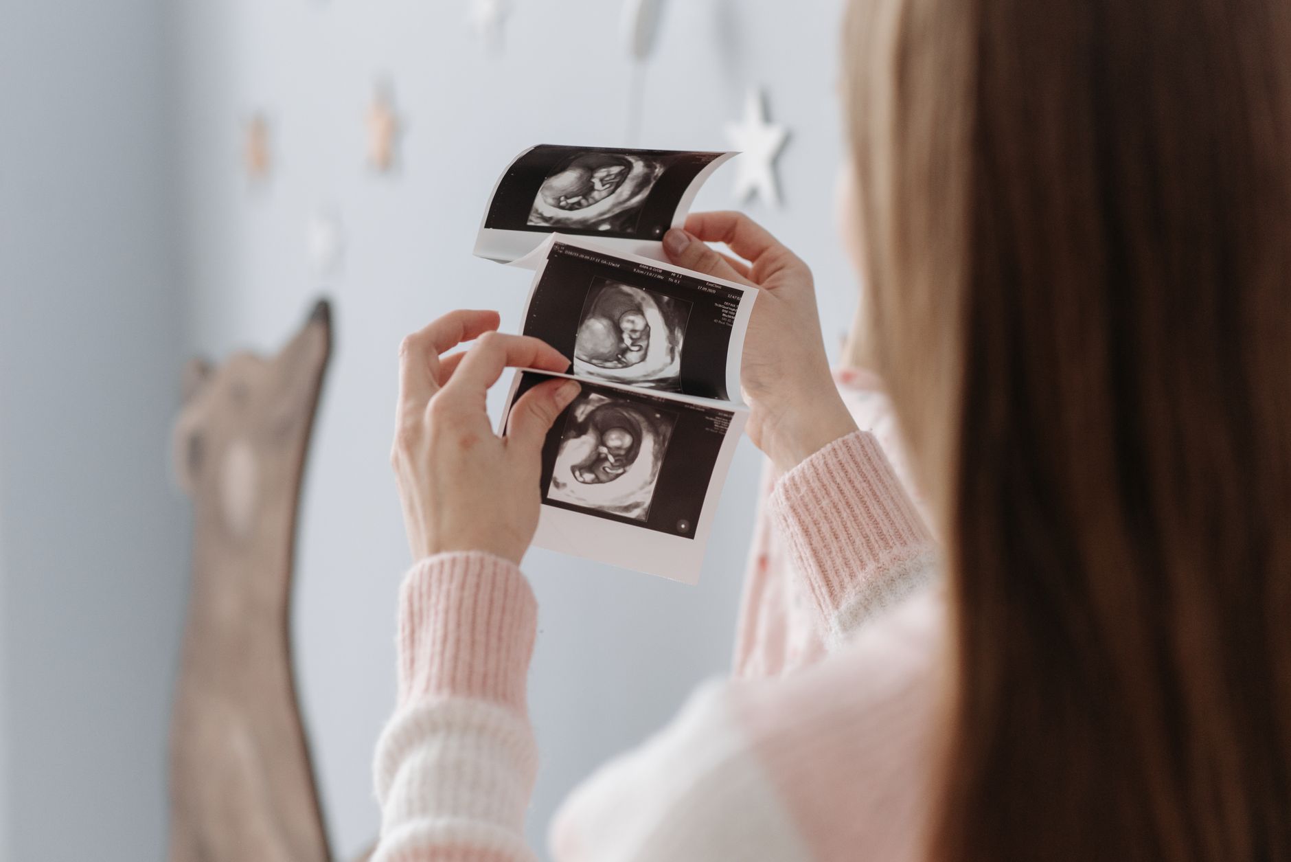
Seeing the tiny heartbeat on the printed image can turn anxiety into joy.
Nutrition tips for 9‑week pregnancy
Early pregnancy is a critical window for building the foundations of fetal development. Focus on nutrient‑dense foods that support rapid cell division and organ formation:
Folate‑rich foods: Leafy greens, lentils, fortified cereals, and citrus fruits help prevent neural tube defects.
Protein: Lean meats, eggs, Greek yogurt, and beans provide the building blocks for tissue growth.
Omega‑3 fatty acids: Fatty fish (low in mercury), chia seeds, and walnuts support brain and eye development.
Iron and vitamin C: Pair iron‑rich foods (red meat, spinach) with vitamin C sources (bell peppers, strawberries) to improve absorption.
Hydration: Aim for at least 8‑10 glasses of water daily to support amniotic fluid production and reduce constipation.
Continue taking a prenatal vitamin that includes at least 400 µg of folic acid, 30 mg of iron, and DHA if possible. Discuss any dietary restrictions with your provider to ensure you’re meeting all nutritional needs.
According to the CDC’s 2023 nutrition guidelines, a well‑balanced diet in the first trimester also helps mitigate common symptoms like fatigue and nausea, making you feel steadier for upcoming appointments.
How many weeks until baby can be seen moving on ultrasound?
Fetal movement, often called “quickening,” is first detectable by ultrasound around 10‑12 weeks. At nine weeks, the embryo’s muscles are just beginning to contract, but the motion is too subtle for most transabdominal scans. By week 12, you’ll typically see small limb wiggles and occasional flickers of the torso, especially on a high‑resolution transvaginal scan.
When you do see movement, it’s a reassuring sign that the nervous system is developing. However, the absence of visible motion at nine weeks is normal and does not indicate a problem as long as the heartbeat is present.
Choose soft, deep‑orange papaya — it’s a tasty source of folate and vitamin C for early pregnancy.
Can you see the baby's heartbeat at 9 weeks?
Yes. By nine weeks most embryos have a strong, regular heartbeat that is easily captured on a standard transabdominal scan. The heart rate typically ranges from 140 to 160 bpm. If the heart is not visible, the sonographer may ask you to fill your bladder more fully or switch to a transvaginal probe for a clearer view.
Why is my 9‑week ultrasound blurry?
Blurriness can stem from a full bladder that hasn’t been filled enough, maternal body habitus, or a low‑lying uterus. Your provider will often recommend drinking 24–32 oz of water 30 minutes before the appointment to create a better acoustic window. If the image remains fuzzy, a transvaginal scan is a safe alternative that usually yields a sharper picture.
Is a transvaginal ultrasound better at 9 weeks?
For many women, especially those with higher BMI or a uterus that hasn’t yet risen out of the pelvis, a transvaginal ultrasound provides superior resolution at nine weeks. It allows the technician to see the embryo’s tiny structures—like limb buds and the yolk sac—more clearly. While it may feel slightly uncomfortable, the probe is small and the procedure is brief. The benefits in diagnostic accuracy often outweigh the minor inconvenience.
Is ultrasound safe for the baby at 9 weeks?
Yes. Ultrasound uses sound waves, not ionizing radiation, and has been used in obstetrics for decades without evidence of harm when performed by trained professionals. The FDA and ACOG state that diagnostic ultrasound is safe when the “as low as reasonably achievable” (ALARA) principle is followed—meaning the shortest possible exposure at the lowest power needed for a clear image.
Most scans last 15–30 minutes, well under the safety thresholds identified in studies. If you have concerns about repeated scans, ask your provider to document the indication and ensure each exam is medically justified.
What to wear and bring to your 9‑week ultrasound appointment?
Comfort is key. Wear a loose‑fitting top that can be easily lifted or a maternity shirt without a bulky bra, because the sonographer may need to access your abdomen. Slip‑on shoes are handy if you need to change after the scan.
Bring a water bottle to stay hydrated, a list of current medications or supplements, and any previous ultrasound images if you have them. Having a notebook for questions (e.g., “What does my CRL mean?”) helps you remember the answers after the appointment.
Doctor’s note
From our medical team: A nine‑week scan is primarily a “viability” check. Seeing a steady heartbeat, an appropriate crown‑rump length, and clear limb buds means your pregnancy is on track. If anything looks atypical, your provider will likely repeat the scan or order additional labs rather than jump to conclusions. Remember, every embryo grows at its own pace, and minor variations are common. Keep taking your prenatal vitamins, stay hydrated, and bring any new symptoms to your next appointment. If you’re ever unsure, a quick call to your midwife or OB‑GYN can provide peace of mind.
Myth vs. fact
Myth: A blurry ultrasound means something is wrong with the baby. Fact: Image quality often depends on maternal factors or the type of probe used. A fuzzy picture is usually resolved by adjusting the technique, not by indicating fetal issues.
Myth: You can find out your baby’s gender at nine weeks. Fact: Gender differentiation typically isn’t possible until the second trimester (around 14‑16 weeks) when the external genitalia have formed.
Myth: A small gestational sac always means a miscarriage is coming. Fact: A slightly small sac can be normal, especially if the embryo’s CRL and heartbeat are appropriate. Your provider will monitor growth rather than assume the worst.
Key takeaways
At nine weeks the embryo is about the size of a grape and shows a clear heartbeat (140–160 bpm).
Key measurements include crown‑rump length (27–31 mm) and gestational sac diameter (20–25 mm).
Both transabdominal and transvaginal ultrasounds are safe; the latter provides sharper images if the first scan is blurry.
Gender isn’t visible yet—wait until 14‑16 weeks for reliable determination.
Typical concerns (small sac, faint heartbeat) often resolve with a repeat scan; persistent issues warrant closer monitoring.
Stay hydrated, maintain a folate‑rich diet, and keep your prenatal appointments to track growth.
Ultrasound is considered safe for the baby when performed by qualified professionals following ALARA guidelines.
Wear comfortable clothing and bring a water bottle to ensure a full bladder for the best image quality.
Frequently asked questions
What does a 9‑week ultrasound look like?
The image shows a tiny embryo within a fluid‑filled gestational sac, a bright yolk sac nearby, and often a flickering dot representing the heartbeat. Limb buds appear as small paddles, and the overall size is comparable to a grape.
Can you see the baby's heartbeat at 9 weeks?
Yes—most nine‑week scans capture a strong heartbeat ranging from 140 to 160 bpm, confirming viability.
How big is the baby at 9 weeks?
The embryo measures roughly 0.6–0.9 inches (1.5–2.3 cm) and weighs about 0.1 gram, similar to a strawberry or grape.
When can you see the baby's gender on an ultrasound?
Gender is usually discernible around 14‑16 weeks when the external genitalia have developed enough for reliable visualization.
Why is my 9‑week ultrasound blurry?
Blurriness often results from a partially filled bladder, higher maternal BMI, or a low‑lying uterus. Drinking more water before the appointment or switching to a transvaginal probe typically clears the image.
Is a transvaginal ultrasound better at 9 weeks?
For many patients, especially those with a higher BMI or a uterus that hasn’t risen out of the pelvis, a transvaginal scan offers higher resolution and clearer views of the embryo and sac.
Do I need to fast before a 9‑week ultrasound?
No. Unlike some abdominal imaging tests, an early‑pregnancy ultrasound does not require fasting. The only preparation needed is a full bladder for the transabdominal portion, which improves sound wave transmission.
Can I have sex after a 9‑week ultrasound?
Yes. After the scan, you can resume normal activities, including intercourse, unless your provider has given you specific medical advice to limit activity for another reason.
When to call your doctor
If you notice any of the following, contact your provider right away: no detectable heartbeat, persistent severe abdominal pain, heavy vaginal bleeding, fever over 100.4 °F (38 °C), or rapid swelling of the hands or face. This information is for educational purposes only and does not replace personalized medical advice.
References
American College of Obstetricians and Gynecologists (ACOG). “Ultrasound in Pregnancy.” 2023 clinical guidance.
National Health Service (NHS). “Ultrasound scan in pregnancy.” Updated 2022.
Royal College of Obstetricians and Gynaecologists (RCOG). “First‑trimester ultrasound.” 2021.
Mayo Clinic. “First trimester ultrasound: What to expect.” 2023.
World Health Organization (WHO). “Recommendations on antenatal care for a positive pregnancy experience.” 2022.
Centers for Disease Control and Prevention (CDC). “Prenatal care: Nutrition.” 2023.
Fetal Medicine Foundation. “Crown‑Rump Length standards.” 2022.
Society for Maternal‑Fetal Medicine (SMFM). “Management of early pregnancy loss.” 2023.
National Institute for Health and Care Excellence (NICE). “Early pregnancy assessment.” 2022.
American Pregnancy Association. “Early pregnancy symptoms and nutrition.” 2023.
U.S. Food and Drug Administration (FDA). “Diagnostic Ultrasound Safety.” 2021.
Society for Radiology (ACR). “ALARA principle for obstetric imaging.” 2020.
Editor's pick for this topic
About the Author
When Shubhra Mishra was expecting her first child in 2016, she was overwhelmed by conflicting food advice — one site said yes, another said never. By the time her second baby arrived in 2019, she realized millions of mothers face the same confusion.
That sparked a five-year journey through clinical nutrition papers, cultural diets, and expert conversations — all leading to BumpBites: a calm, compassionate space where science meets everyday motherhood.
Her long-term vision is to build a global community ensuring safe, supported, and free deliveriesfor every mother — because no woman should face pregnancy alone or uninformed. 🌿
🌍 Stand with mothers, shape safer guidance
Join a small circle of experts who review BumpBites articles so expecting parents everywhere can decide with confidence.